

Breast Ultrasound Scan near Wolverhampton
Private Breast Ultrasound Scans near Wolverhampton
Breast Scans near Wolverhampton
A private ultrasound breast scan near Wolverhampton is a non-invasive and painless imaging test that uses sound waves to create pictures of the breast tissue. It can help detect abnormalities in the breast, armpits or axilla, cysts, lumps, masses or infections, and evaluate the blood flow to the breast area.
Some reasons why someone would need a private ultrasound breast scan are
- Symptoms such as breast pain, swelling, nipple discharge, skin changes, sunken nipples, changes in shape or size, fluid excretion, skin sores, lumps and bumps, dimples, red or hot areas, and nipple crust.
- Dense breast tissue reduces the visibility of tumours in mammograms.
- Breast implant problems may make it difficult to detect breast cancer on a mammogram.
- Personal or family history of breast cancer.
- A need for a second opinion, or after a home self-check.
- Breast Health Check before surgery or post-surgery
- Breast symptoms for people who cannot have mammograms, pregnant women or younger women (under 50 who are usually not routinely invited for NHS breast screening, unless they have risk factors or have a close family member with young onset of breast cancer)

Our Breast Ultrasound Scan
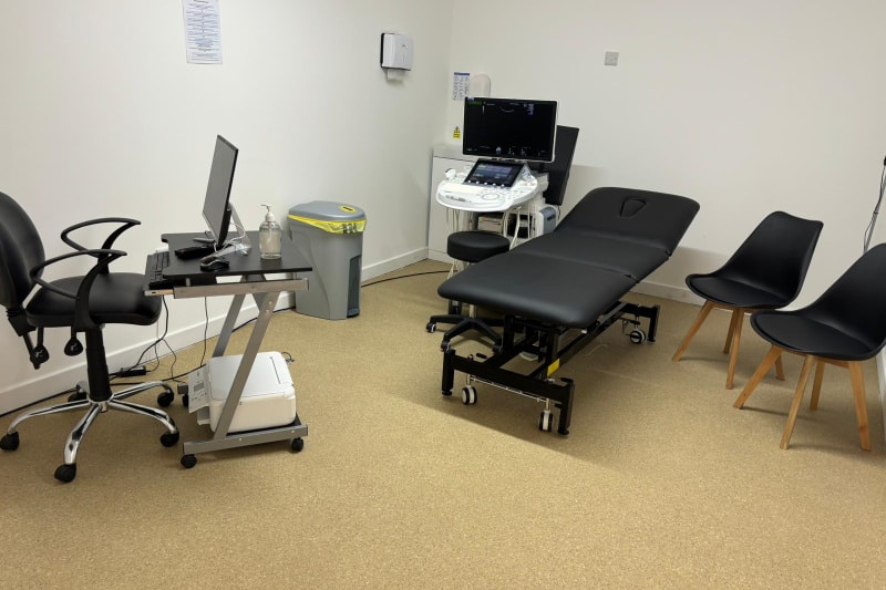
At Universal Ultrasound Diagnostics, we are dedicated to providing exceptional private breast ultrasound scans at our breast ultrasound clinic in Birmingham, UK. We offer flexible and convenient dates and times that suit your needs and preferences.
With our cutting-edge Ultrasound systems and highly skilled, qualified and experienced staff, we deliver precise and reliable imaging for both our private clients and referring practitioners in a comfortable and discreet setting.
Our commitment to utilising the latest technology ensures we consistently deliver the highest quality performance you can trust.
Breast Scan Timeline & Cost
Typical Cost
You can see our latest rates on our booking page.
Scan Time
Breast ultrasound scans may take up to 30 to 50 minutes, depending on the circumstances.
Results
The sonographer will discuss any immediate findings with you, and the report and images will be emailed to you on the same day.
SAME-DAY PEACE OF MIND FOR BREAST SCANS
Health and peace of mind should never be delayed. Our Same-Day Breast Scans are designed to provide swift, quality service, answer your questions, give you peace of mind, and ensure you do not have to endure unnecessary uncertainty or waiting. Whether it is an urgent concern or a routine check, we prioritise your well-being with our state-of-the-art technology, highly qualified and trained staff with vast experience, and compassionate care.
We discuss any findings after your scan and make recommendations for further clinical correlation, where needed. When it comes to your health, every minute matters.

How good is ultrasound at detecting breast cancers ?
For imaging palpable abnormalities in the breast, a breast ultrasound has been proven to be an exceptionally effective tool. The efficiency in detecting breast cancer is dependent on various factors, such as the type of breast tissue, the sonographer’s or radiologist’s skills in scanning, and the ability to interpret the images of the lesion, tumour, or mass characteristics.
Breast ultrasound is usually excellent for people with dense breasts, as an ultrasound can detect cancers that a mammography may miss. However, mammography can pick up small calcium deposits known as microcalcifications, which ultrasound may miss.
The ultrasound can distinguish between cystic lesions (fluid-filled cysts) and solid lumps. It can reveal the features of a solid lump or mass that would suggest that the lump or mass is malignant, indeterminate, or suspicious.
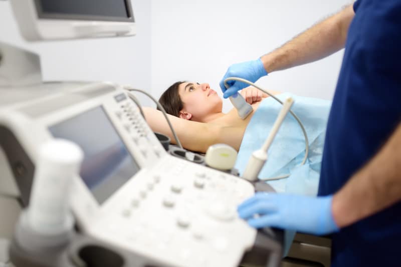
A Breast Ultrasound near Wolverhampton is mainly utilised for targeted assessment of palpable lumps or as a supplementary tool to MRI or mammography. It is not normally used as a primary screening tool except in pregnant women, younger women, or people who cannot have mammograms.
Checking your breasts regularly is one of the simplest ways to stay breast aware.
A quick self-check helps you learn what's normal for you, so you're more likely to notice a change early, whether that's a new lump, a thickened area, or a change in the skin or nipple. Most breast changes are benign and can be linked to hormones, your menstrual cycle, pregnancy, breastfeeding, or natural changes with age. Still, it's always worth taking anything new or unusual seriously and getting it assessed.
A helpful way to remember what to do is TLC: Touch, Look, Check.
Touch: Using the pads of your three middle fingers, move in small circles and work methodically across the whole breast, right up to the collarbone and into the armpit area. Use light, medium, then firmer pressure so you can feel different depths of tissue. Take your time and follow a consistent pattern (circular, up-and-down, or wedge sections).
Look: Stand in front of a mirror with your arms by your sides, then hands on hips, then arms raised. Look for changes in shape, size, or contour, dimpling or puckering of the skin, redness, crusting, new nipple inversion, or any discharge that happens without squeezing.
Check positioning: You can examine lying down, standing, or in the shower. Aim for once a month, ideally at the same point in your cycle when your breasts are least tender.

If you do find a lump or change, a private breast ultrasound can be a helpful next step. A breast ultrasound scan near Wolverhampton can often tell whether a lump looks fluid-filled (a cyst) or solid, and it can also assess surrounding tissue and lymph nodes. If you're concerned, always speak to your GP, and if something keeps changing even after a normal scan, it's important to get it reviewed again.
For a more detailed, step-by-step guide, read our full blog: Private Breast Screening: How to Check Your Breasts.
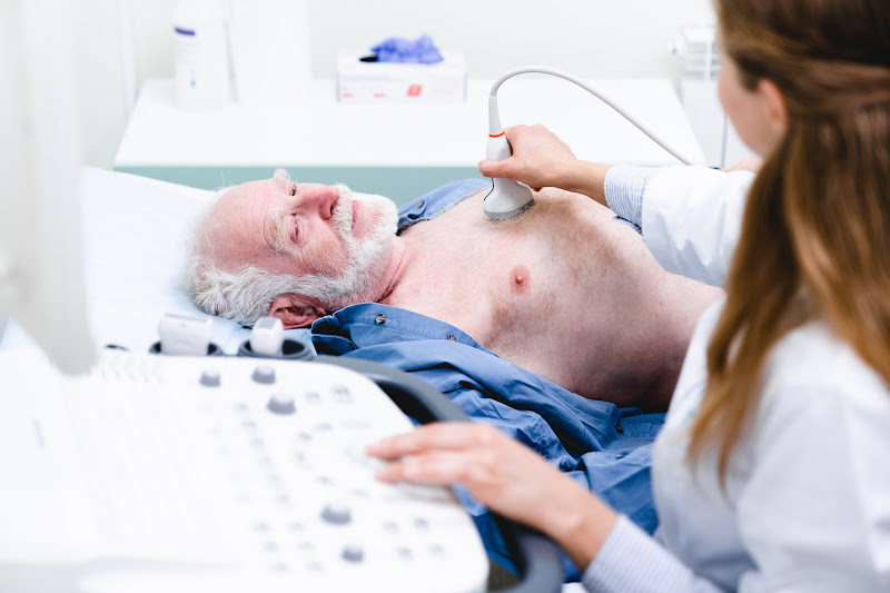
Breast Ultrasound Scans for Men Wolverhampton
The majority of male breast issues are considered benign; however, it is recommended that male breasts should still be properly evaluated to rule out anything that could potentially be serious, especially if you have symptoms.
The most common cause of male breast enlargement remains gynaecomastia, which can be linked to medication, underlying health conditions, changes in weight, hormonal changes, or even the use of steroids. It can present as a firm or soft swelling beneath a nipple, affecting one breast or both. Whilst cancer is extremely rare in male breasts, it can still occur. Other breast problems include infection, cysts, and fatty tissue (lipomas).
Symptoms
Some symptoms are frequently overlooked, which can inturn delay diagnosis; early detection is always critical if there is an issue of concern.
Signs to look out for include:
- A growing or newly developed lump within the breast or chest wall
- Nipple discharge, inversion or any other unusual changes
- Persistent breast pain
- Changes to the breast skin, which might include redness or dimpling
How Ultrasound Helps
A breast ultrasound scan is one of the first imaging techniques used to assess the breast.
The sonographer will assess the breast during the scan, and this includes
- The appearance - shape, size, and characteristics of any lump or cyst
- The chest wall and the surrounding breast tissue
- May also assess the armpit/axilla
- Whether a lump is solid or fluid-filled
- Checking the blood flow where needed
This detailed examination helps to distinguish between features that may require further testing or investigation and benign conditions. In most cases, a breast ultrasound scan can correctly detect benign causes such as a simple cyst or gynaecomastia, giving you peace of mind.

Why Choose Our Private Ultrasound Scan Clinic?
1. Expert Sonographers: Our clinic boasts highly skilled and experienced sonographers specialising in Diagnostics and Pregnancy scans.
2. State-of-the-Art Technology: We use cutting-edge ultrasound systems for precise and reliable imaging.
3. Comfortable and Discreet Setting: Our clinic ensures a comfortable and confidential patient environment.
4. Flexible Appointments: We offer flexible dates and times to suit your convenience.
5. Prompt Results: Immediate findings are discussed during your appointment, and a detailed report and images are emailed to you the same day.
Meet the Team
Lydia, our CQC Registered Manager and Operations Director, leads the team with empathy, faith and the utmost integrity. Her patient-focused approach helps to shape the culture of our service.
Our Consultant Radiologist Dr Moti, is supported by our advanced sonographer Andrew, combine clinical excellence with reassurance, giving our patients peace of mind and genuine warmth. Ali brings clarity to every scan and calm assurance, helping patients feel at ease, supported, and well-informed.
Behind the scenes, Emma, our practice manager, plays a pivotal role, ensuring every detail of the patients’ journey is seamless. She works alongside the Registered Manager to ensure quality assurance is maintained.
Carly, our Medical Administrative Officer, plays a crucial role in patient experience and works alongside the Registered Manager to maintain high-quality standards through promoting continuous improvement across the practice.
Together as a team, we are united by one purpose:
Our Mission - To deliver high-quality care with compassion that not only meets the patients’ expectations but goes over and beyond to exceed expectations.
Our Mission, Vision And Values
Mission - To be the most efficient and preferred diagnostic imaging centre that provides excellent quality care, that not only meets but goes above and beyond to exceed patients’ expectations.
Vision - Our vision is innovation as healthcare and technology are continuously evolving, which will enhance scanning techniques, image quality, accuracy, and efficiency and provide clearer insights into diseases.
We are proud to be a compassionate, caring, and respectful team. We do this by asking staff to work within our core values:

Compassionate by being inclusive, attentive, caring, respectful and showing kindness to staff, our patients, and their families.

Caring by going beyond compliance, performing above and beyond to improve our services and providing person-centred care by involving patients in their care, offering choices, and promoting dignity and privacy.

Respectful by active listening, seeking to understand others, showing empathy for differences, apologising when things go wrong, being polite and showing gratitude to our staff and patients.
It's easy to book an appointment.
Universal Ultrasound Diagnostics offers an online booking system for easy appointment scheduling. There are options for walk-ins depending on availability: urgent scans within 5 days and routine scans within 14 days.
Appointment notifications and reminders are sent via email. The system supports self-check-in and online referrals by GPs.
We provide chaperones and interpreting services and allow family presence during pregnancy scans.
Our facility is accessible by public transport and has parking.
We allow family presence for most scans; however, children are not allowed in the room for certain scans. Please check your appointment confirmation email for clarity on our safety policy.
Our FAQ page answers a lot of general questions about your scan.
There is parking around The Green or Pay & Display at the side of The Bulls Head Pub, which charges £1 per hour (cash and card accepted). We are open 8 am to 11 pm Monday to Sunday.
Opening Times
Monday to Sunday 8 am - 11 pm
Location
10c The Green, Kings Norton, Birmingham, B38 8SD

Breast Scan Faqs
What is a breast ultrasound scan?
Breast ultrasound scans are a type of scan used to look at the soft tissue of the breast. Ultrasound uses high-frequency sound waves to create a 2D image of soft tissues and organs within the body and is much safer than X-rays as it does not use radiation.
Schedule an appointment for a private ultrasound breast scan at our clinic near Wolverhampton
Symptoms of breast issues?
Symptoms of breast issues
- Bumps
- Dimples
- Nipple crust
- Growing veins
- Thick areas of skin
- Sunken nipples
- Red or hot areas
- New shape/size
- “Orange peel” skin
- Fluid excretion
- Skin sores
- Hard lumps
Get Same-day peace of mind with a private ultrasound scan near Wolverhampton
Causes of breast issues?
Causes of breast issues include:
• Tumour
• Cyst
• Infection
• Fat necrosis
• Fibroadenomas
Get Same-day peace of mind with a private ultrasound scan near Wolverhampton
What is a tumour?
A tumour is a solid mass of tissue caused by a collection of abnormal cells in one area. Tumours can be both benign and malignant. An ultrasound will be able to detect the presence of a tumour however it will not be able to differentiate between the types.
What is a breast cyst?
A breast cyst is a fluid-filled sac that develops in the tissue of the breast. They commonly occur in women between 30 to 50 years old however, these can develop at any age as they are related to hormonal fluctuations. A breast cyst is part of a condition known as fibrocystic breast changes and they are usually benign (non-cancerous).
Causes
Commonly linked to hormonal changes, especially oestrogen. The exact cause of a breast cyst is not always clear; however, they can develop if glands in the breast enlarge or start to produce excess fluid.
Symptoms
- A lump that can be felt in one or both breasts (this may feel firm, smooth, or round).
- Tenderness or swelling in the breast.
- Discomfort or pain around the lump (which may worsen before menstruation).
- Sometimes cysts are too small to be felt and are incidentally detected through imaging such ultrasounds.
TYPES OF BREAST CYSTS
1. Simple Cysts:
- Filled only with fluid.
- Smooth, round, and easy to differentiate from other breast lumps.
- Generally benign (non-cancerous)
2. Complex Cysts:
- Contain both fluid and some solid components (like debris).
- May require additional tests (such as biopsy) to rule out the possibility of malignancy (cancer).
3. Microcysts:
- Tiny cysts that are not large enough to feel during a physical exam.
- These are detected only through the use of medical imaging, such as an ultrasound scan.
4. Macro Cysts:
- These are large cysts that are usually easily felt and may even be visible to the naked eye. They can cause discomfort or pai due to their size.
- These sometimes press on surrounding tissues and may lead to other problems.
TREATMENT PLANS FOR BREAST CYSTS
1. For Simple Cysts:
- No Treatment Necessary: If they are small, painless, and not causing concern it is common that there will be no further action required.
- Aspiration: Fluid may be removed with a thin needle to alleviate any pain or discomfort experienced.
- Regular Monitoring: Periodic imaging to check for changes may be requested, especially if the cysts are recurring.
2. For Complex Cysts:
- Biopsy: If there are solid components to the cyst, a biopsy may be
performed to ensure there is no malignancy (cancer). - Surgical Removal: Rare but may be recommended if features are
suspicious/malignant or if the cyst persists after drainage.
3. For Symptomatic Macro Cysts:
- Fluid Aspiration: To reduce pressure and pain by draining fluid.
- Lifestyle Adjustments: Wearing supportive and correctly fitted bras,
reducing caffeine intake, and applying warm compresses to the area may
help to ease discomfort.
4. For Hormonal-Related Cysts:
- Hormonal Therapy: In some rare cases, birth control or other hormone
regulation medicine may be used to decrease hormonal fluctuations that
are the cause of reoccurring cysts.
WHEN TO SEEK MEDICAL ATTENTION
If a cyst has show unusually rapid growth, becomes increasingly painful, or has other worrying characteristics such as blood or fluid coming from the area then further evaluation is necessary. Attending routine mammogram appointments and carrying out regular breast self-examinations can help to monitor any changes.
DIAGNOSIS
Medical professionals typically diagnose breast cysts through:
- Clinical Examination: Palpating (gently pressing) the lump to assess its features such as appearance and hardness.
- Imaging Tests: Mammograms and ultrasound scans help identify cysts. However, an ultrasound is a better modality that is often used to further analyse a lump or mass and help to determine if it is a solid mass or cyst.
- Fine Needle Aspiration: Using a thin needle to extract fluid, which can be used to confirm the presence of malignant/cancerous cells (cytology) or may be used to improve symptoms.
Self-examinations are also an important part of any self care regime.
TREATMENT
Most breast cysts do not require treatment unless they cause pain or discomfort:
- Observation: You can monitor the cysts; however, small, painless cysts often resolve on their own.
- Aspiration: For larger cysts, draining the fluid can sometimes be necessary, especially to relieve symptoms, your GP may recommend this.
Surgical Removal: This is rare and is usually only considered if the cyst is persistent or shows concerning or unusual features.
PREVENTION & MANAGEMENT
It is not always possible to prevent breast cysts from occurring, however, maintaining a healthy lifestyle can help to manage hormonal fluctuations. Wearing a supportive and well-fitted bra, reducing caffeine intake, and managing stress can also help to alleviate symptoms in some people.
Get Same-day peace of mind with a private ultrasound scan near Wolverhampton
What is fat necrosis?
Fat necrosis is the death of the adipose tissue located in the breast. It most commonly develops after an injury or trauma to the area but is usually a benign condition. Fat necrosis can cause a lump to form which may need to be tested with a biopsy.
What is fibroadenoma?
A fibroadenoma is a solid breast lump that most commonly occurs between the ages of fifteen and thirty-five but can be found in anyone who has periods. It is a small, smooth, rubbery lump that moves easily within the breast tissue. They are common and benign lumps, however they should be monitored for any sudden change in size or feel.
Is a breast ultrasound scan better than a mammogram
Ultrasounds and mammograms each have various weaknesses and strengths. These two primary imaging modalities are widely used for the screening and diagnosis of breast conditions. Ultrasound is usually considered safe, painless, non-invasive, and fast. High sound waves are used to obtain breast images with the help of an ultrasound machine and mammography is a form of low-dose x-ray imaging.
Breast ultrasound is often used for women under 45, and pregnant women and it is a primary investigation in younger women in detecting breast problems. It also has the advantage of detecting lesions in dense tissue of young women where the accuracy of mammography is sometimes limited due to overlying structures. Mammography may obscure lesions in dense tissue or scar tissue but provides better information when large breast tissue is visualised.
Ultrasound has a significant role, especially in the assessment of masses in older women. However, age should not be considered as a limiting factor in deciding to have either an ultrasound or mammography. Individual circumstances, including your age, breast density, family history, and symptoms should be considered to enable an informed decision regarding breast investigation. Sometimes mammography may also be recommended alongside ultrasound for detailed and comprehensive evaluation.
Whilst ultrasound is a safer technique that does not use radiation, small deposits of calcium in the breast called microcalcification may be missed on ultrasound. Mammography would be the most effective for the detection of these tiny deposits. These deposits may be early indicators of breast cancer. Ultrasound is much less effective as it cannot properly visualise the entire breast, or penetrate deep breast tissues, or the axillary lymph nodes. Further investigations are helpful in these scenarios.
Get in touch with Universal Ultrasound Diagnostics, for a Breast Scan near Wolverhampton
Why would you be referred for an ultrasound after a mammogram?
A breast ultrasound primarily focuses on resolving concerns identified during a mammogram or a physical examination of the breast. It serves as a valuable diagnostic tool to discern whether a breast abnormality is a benign cyst filled with fluid or a solid tumour.
Breast ultrasounds can be performed as additional tests after a mammogram or to complement as part of further investigations. For instance, where your mammogram has revealed a lesion on your breast, an ultrasound may be requested to help determine if it is a solid mass or cyst. Ultrasound provides detailed information on the lesion/lump while mammography helps to visualise early changes, asymmetry, and concentrated tissue that may be due to some growth.
Breast ultrasound is particularly beneficial in specific scenarios, including:
- Dense Breast Tissue: If you have dense breast tissue then ultrasound would provide more information and detailed images as mammography has limitations in this regard because lesions may be underdiagnosed.
- Fluid-filled/Solid lesions: Ultrasound is significant for the detection of fluid-filled lesions (cysts) and solid tumours.
- Age below 25: As the risk of cancer is relatively lower in this age group, ultrasound may be preferred.
- Role in pregnancy: Mammography uses X-ray radiation which may be hazardous for the pregnant individual and the baby in the mother’s womb so ultrasound may be a choice of investigation and favourable for ensuring the safety of the mother and the baby and in helping early detection of disease.
- Breast health check before breast implants. The scan can help detect any abnormalities or problems
- Checking for any breast implant leakages. Ultrasound can identify holes, tears, and leakages of silicone gel or saline in the breast tissue.
Your GP may help you to decide the best investigation for you particularly when you have a palpable lump, symptoms, or family history that is positive.
Early detection of the disease especially early-stage cancer leads to better outcomes. Hence self-examination, routine and regular breast screening are recommended by the doctors.
Interested in a private Breast Scan near Wolverhampton, contact Universal Ultrasound Diagnostics here
We also offer blood tests relating to the breast
• Carcinoembryonic Antigen (CEA)
• Cancer Antigen 125 (CA-125)
What kinds of things can breast ultrasounds detect?
As stated above, a breast ultrasound scan can detect tumours, cysts and infections. Though ultrasound scans alone cannot be used to diagnose disease and must be used in conjunction with other diagnostic tests.
What do I wear for a breast scan?
You will need to wear a loose fitting top that can easily be opened or removed so that the sonographer can access the breast. Where possible the sonographer will only have you uncover the breast that is being scanned and every effort will be made to protect your dignity during the scan.
Do I need to consent to a breast scan?
All patients must give their consent (permission) before undergoing an examination. If they are unable to give consent themselves then a person with power of attorney will need to give consent on their behalf. Please ensure that the pre-treatment consent form is completed via the embedded link in your confirmation email before you attend the appointment. The scan will not go ahead if this form is not completed.
Are breast scans painful?
Breast scans are not painful but they may be uncomfortable as the sonographer will have to gently press the ultrasound scan probe to your breast. They may also ask you to change position, such as lying on your side after lying on your back, to check the tissue thoroughly. They may also request to check the lymph nodes in your armpits.
What are the red and blue blobs on the ultrasound?
During the scan the sonographer may use a colour doppler to check blood flow in your tissue and organs. This will appear as blue and red blobs on the image and are nothing to be concerned about.
What happens during the scan?
The sonographer will put ultrasound gel onto your skin and move a probe over your breast. They will examine each part of your breast multiple times and in different positions to check for any areas of concern. They may also ask to check your armpits as this is where your lymph nodes are located. Do not be alarmed if the sonographer checks the same area again or goes quiet during the exam. They will explain any findings to you in detail once the scan is complete.
What is the next step after a breast ultrasound?
After a breast ultrasound, the next steps depend on the scan findings. Below are some possibilities:
Normal Results: If the scan appears normal, no further tests may be required
- Fluid-Filled Lump: If a fluid-filled sac or lump is detected, a doctor may decide to drain the fluid. Doctors may drain a fluid-filled lump in the breast for various reasons. Breast cysts, typically noncancerous, are fluid-filled sacs that often feel like water-filled balloons or grapes. If a cyst becomes painful, large, or uncomfortable, draining the fluid can relieve symptoms. This procedure involves using a thin needle to extract the fluid. Surgery to remove the cysts might be considered if the fluid recurs frequently. It is crucial to monitor any changes in your breasts and seek medical attention if you notice any changes in the skin or new lumps.
- Solid Lump: Additional tests may be required based on the scan findings if a solid lump is detected. Your doctor may recommend a biopsy, blood test, mammogram, or breast MRI.
Your doctor will further explain these in-depth, provide guidance based on the ultrasound results, and recommend any necessary follow-up procedures. If you have any concerns, reach out to your doctor.
What is a breast implant ultrasound scan?
A breast implant ultrasound scan is a safe and painless examination that does not use radiation. It uses high-frequency sound waves to produce detailed images of breast tissues and the surrounding areas to help identify any leakages, ruptures of the implants, verify the implant’s location or for any unusual changes or fluid accumulations in the breast.
What is a Breast Implant?
A breast implant is a procedure where a soft, silicone shell filled with silicone gel or saline (sterile salt water) is surgically positioned beneath the breast tissue and chest muscles to either reconstruct the breast following a lumpectomy or injury (medical purposes) or to increase the breast’s size (cosmetic reasons).
Why is it important to monitor the integrity of breast implants?
Breast implants may deteriorate or burst over time. These ruptures for silicone implants may be “silent,” which means they do not exhibit any signs. If a leak or rupture is not detected, the silicone gel has the potential to spread and result in lumps or irritation. The implant may stiffen (capsular contracture) or change form leading to discomfort or health hazards. Monitoring guarantees the early identification of problems before they worsen, enabling prompt replacement or treatment.
When should a breast ultrasound be used to monitor breast implants?
An ultrasound scan of the breast to check implants may be required when there are changes in shape, feel a lump, stiffness, discomfort, pain, if the implant rupture is suspected, or as a health check after breast implant surgery. A routine ultrasound to monitor breast implants is recommended for five to six years following silicone implant placement, and then every two to three years after that. An ultrasound can be used as the first modality to check any issues with breast implants, as it is less expensive, faster, and still effective.
Looking to check the integrity of your breast implants? Book your reassurance ultrasound scan near Wolverhampton today.