

Fertility Ultrasound Scan Walsall
Private Fertility Ultrasound Pelvic Examinations at our Birmingham Clinic near Walsall
Fertility Ultrasound Scan Near Walsall
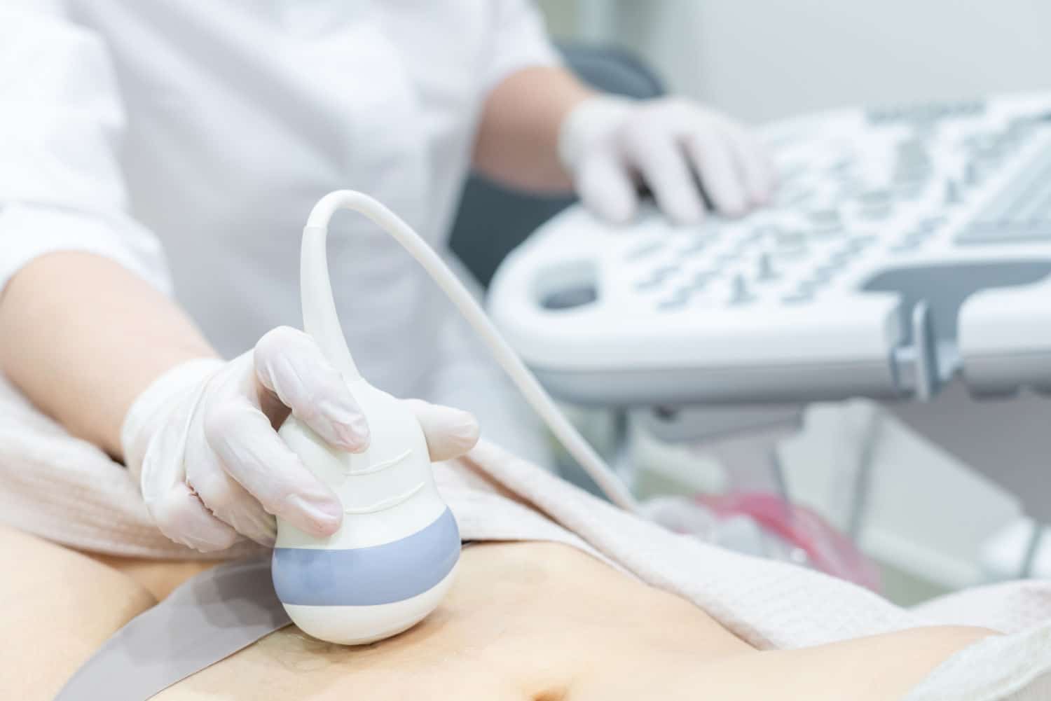
A fertility ultrasound scan is a pelvic examination that utilises high-frequency soundwaves to create images of the inside of your body. The scan is used to monitor your cycle, check how treatment is proceeding, and when to retrieve and implant eggs.
A fertility scan is used to assess the overall state of your uterus, endometrium, ovaries, and fallopian tubes.
Fertility Scan Time & Cost
Typical Cost
You can see our latest rates on our booking page.
Scan Time
Scans can take 30 - 50 minutes.
Results
The sonographer will discuss any immediate findings with you, and the report and images will be emailed to you on the same day.
Our Fertility Ultrasound Scans
At Universal Ultrasound Diagnostics, we are dedicated to providing exceptional private fertility ultrasound scans at our diagnostics ultrasound clinic in Birmingham, UK. We offer flexible and convenient dates and times that suit your needs and preferences. With our cutting-edge Ultrasound systems and highly skilled, qualified and experienced staff, we deliver precise and reliable imaging for both our private clients and referring practitioners in a comfortable and discreet setting. Our commitment to utilising the latest technology ensures we consistently deliver the highest quality performance you can trust.
About your Fertility Ultrasound Scan appointment
Preparation to do before your scan appointment
You must attend with a full bladder. It is advisable to drink about 1½ to 2 pints of clear fluid an hour prior to the appointment. You may drink water or other non-fizzy drinks, black tea or coffee, or fruit juice. When your bladder is correctly filled you will feel the need to urinate. If it becomes painful, it is too full, and you may partially empty if you can.
If you are unsure, please contact us on 0121 392 3112 or your doctor for further advice.
Please arrive 5 to 10 minutes before your actual appointment time as it may not be possible to scan you if you are late, resulting in your payment being forfeited as per our payment and refund policy.
ARRIVING FOR YOUR FERTILITY ULTRASOUND SCAN APPOINTMEN

The scan is completed in two parts. You will need a full bladder for the first part which is the transabdominal. The sonographer will put ultrasound gel on your skin over your lower abdomen then move an ultrasound transducer slowly over your skin. This will help us to produce better-quality images.
After this is completed, you will be asked to go to the toilet and relieve yourself then return for the second part of the scan which is internal through the vagina. We do not perform transvaginal (internal) examinations on children or persons who have never been sexually active. If you have never been sexually active and do not wish to have an internal examination, please notify the sonographer.
The internal part of the scan gives the most detailed images. While a patient has the right to decline the internal part of the scan this will limit what the sonographer is able to observe and comment on. A chaperone will always be present in the scan room. We will ensure your dignity is maintained throughout your scan.
During the examination
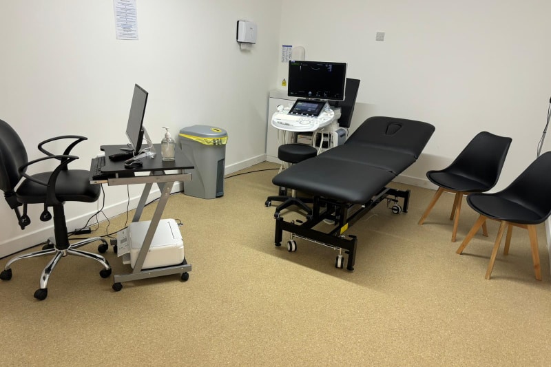
You will be asked to lie on a couch in the examination room. The sonographer performing the scan will ask you to expose your lower abdomen. The gel will be applied over your abdomen, and it may feel cold. The sonographer will gently move an ultrasound probe over your lower abdomen to obtain images of the pelvis. Unless the area being examined is tender, the examination is not painful.
For better visualisation of your uterus and ovaries, a transvaginal scan will be performed with your consent. This can still be performed even during your period. After the transabdominal scan, you will be asked to empty your bladder and return to the ultrasound examination room.
You will be asked to take off your pants and lie down on a couch on top of a wedge cushion. The sonographer will ensure your lower body is covered with a sheet at all times to maintain your dignity. A chaperone will always be available in the scan room. Your decision/choice not to have a chaperone will be respected.
An ultrasound probe will be covered with a disposable latex or non-latex sheath and lubricated with gel. (Please let the sonographer know if you are allergic to latex). The probe will then be placed into the vagina to obtain images of the pelvic area, particularly the ovaries and uterus. The exam may be uncomfortable but not painful.
We do not perform transvaginal examinations on children or on persons who have never been sexually active. If you do not wish to have an internal examination performed, you can discuss this with the sonographer when you come for your scan.
Your decision/choice will be respected. However, there will be limitations to the quality of images obtained without performing a transvaginal examination. Both parts of the examination may take up to 20 minutes, depending on individual circumstances.
AFTER THE EXAMINATION
The sonographer will discuss any immediate findings with you, and the report will be emailed to you on the sameday. An option to receive images is available for a small charge.

Why Choose Our Private Ultrasound Scan Clinic?
1. Expert Sonographers: Our clinic boasts highly skilled and experienced sonographers specialising in Diagnostics and Pregnancy scans.
2. State-of-the-Art Technology: We use cutting-edge ultrasound systems for precise and reliable imaging.
3. Comfortable and Discreet Setting: Our clinic ensures a comfortable and confidential patient environment.
4. Flexible Appointments: We offer flexible dates and times to suit your convenience.
5. Prompt Results: Immediate findings are discussed during your appointment, and a detailed report and images are emailed to you the same day.
Our Mission, Vision And Values
Mission - To be the most efficient and preferred diagnostic imaging centre that provides excellent quality care, that not only meets but goes above and beyond to exceed patients’ expectations.
Vision - Our vision is innovation as healthcare and technology are continuously evolving, which will enhance scanning techniques, image quality, accuracy, and efficiency and provide clearer insights into diseases.
We are proud to be a compassionate, caring, and respectful team. We do this by asking staff to work within our core values:

Compassionate by being inclusive, attentive, caring, respectful and showing kindness to staff, our patients, and their families.

Caring by going beyond compliance, performing above and beyond to improve our services and providing person-centred care by involving patients in their care, offering choices, and promoting dignity and privacy.

Respectful by active listening, seeking to understand others, showing empathy for differences, apologising when things go wrong, being polite and showing gratitude to our staff and patients.
It's easy to book an appointment.
Universal Ultrasound Diagnostics offers an online booking system for easy appointment scheduling. There are options for walk-ins depending on availability: urgent scans within 5 days and routine scans within 14 days.
Appointment notifications and reminders are sent via email. The system supports self-check-in and online referrals by GPs.
We provide chaperones and interpreting services and allow family presence during pregnancy scans.
Our facility is accessible by public transport and has parking.
We allow family presence for most scans; however, children are not allowed in the room for certain scans. Please check your appointment confirmation email for clarity on our safety policy.
Our FAQ page answers a lot of general questions about your scan.
There is parking around The Green or Pay & Display at the side of The Bulls Head Pub, which charges £1 per hour (cash and card accepted). We are open 8 am to 11 pm Monday to Sunday.
Opening Times
Monday to Sunday 8 am - 11 pm
Location
10c The Green, Kings Norton, Birmingham, B38 8SD

Fertility Ultrasound Scan Faqs
Is a Fertility Ultrasound Scan safe?
A fertility ultrasound scan is safe and does not have any of the risks associated with ionising radiation. It has been used to provide valuable images and other diagnostic information with no evidence of any harm. However, as a precaution, the sonographer will take every care to ensure that the level and duration of the ultrasound examination are not more than necessary to obtain the diagnostic images or information to minimise any potential risk to the patient.
Schedule a Fertility Ultrasound Scan at our Clinic near Walsall
What is a Fertility Ultrasound scan?
A fertility ultrasound scan is a pelvic examination that utilises high-frequency soundwaves to create images of the inside of your body. The scan is used to monitor your cycle, check how treatment is proceeding, and when to retrieve and implant eggs.
What are some of the symptoms of infertility?
Infertility can present in various ways, depending on the individual and the underlying cause. Some common symptoms of infertility include:
• Painful intercourse or periods
• Difficulty conceiving after one year of unprotected intercourse
• Abnormal sperm count or quality in men
• Irregular menstrual cycles or no periods
Can you see infertility on an ultrasound?
A fertility ultrasound is performed on your lower abdomen, to assess the overall condition of your ovaries, uterus, and fallopian tubes.
Uterus
The uterus or womb where a baby develops, is examined for the following:
Uterine size and shape: Abnormal shapes, such as a bicornuate (heart-shaped) uterus or a uterine septum (partitioned uterus), can impede implantation or provide inadequate space for the fetus.
- Endometrium Thickness: The lining of the uterus should be the correct thickness. If it is too thick, there may be a hormonal issue. If it is too thin, embryos are less likely to implant. The lining's thickness varies throughout the menstrual cycle, therefore knowing your cycle stage may help in determining if the thickness is appropriate at that stage of the cycle.
- Polyps: Cervical or Endometrial polyps, can grow extending into the uterine cavity, this can prevent embryo implantation similar to a contraceptive coil.
- Fibroids: Large fibroids, which are muscle tissue balls, can alter the shape of the uterine cavity, causing implantation issues or insufficient space for fetal growth.
- Blood Flow: Doppler imaging, which measures blood flow using sound wave frequency changes, is often used in private fertility assessments but is not standard in national health services. Good blood supply to the uterus and its lining is crucial for transporting oxygen and nutrients and for proper lining development
Ovaries
The ovaries, where the eggs are stored, are examined for:
- Endometriosis Cysts: Endometriosis, where the uterine lining is found outside the uterus, can cause cysts that interfere with egg maturation and release.
- Multiple Cysts: This could indicate Polycystic Ovary Syndrome (PCOS), which requires further blood tests for confirmation.
- Other Cysts: Large cysts can disrupt ovarian function, affect egg release, and may cause pain
- Antral Follicles: The follicles are measured and counted to estimate fertility potential, typically in private fertility assessments
- Blood Flow: Good blood flow to the ovaries ensures a sufficient supply of oxygen and nutrients. Doppler imaging is used for this assessment in private fertility settings, often alongside an antral follicle count,
Fallopian Tubes
The fallopian tubes which connect the ovaries to the uterus, are examined for:
- Endometriosis Signs: While endometriosis is difficult to detect on the fallopian tubes, kinking or scarring can indicate its presence, potentially blocking the tubes or affecting cilia which are tiny hair-like structures that aid egg movement.
- Swelling: This may indicate a blockage, however, this can be confirmed by a Hysterosalpingogram (HSG) if detected.
- Scarring: This can result from endometriosis or infection, potentially blocking the tubes or interfering with cilia movement.
Monitoring any changes in your reproductive system and seeking medical attention if you notice new lumps or skin changes is essential.
What are some of the causes of infertility?
Infertility can be caused by a variety of factors, including:
• Chronic stress and emotional trauma
• Hormonal imbalances
• Genetic predisposition
• Nutritional deficiencies
• Environmental toxins
• Side effects of medications or treatments such as chemotherapy
• Medical conditions such as polycystic ovary syndrome (PCOS) or endometriosis
If you have any concerns, you need to discuss this with your doctor who can advise specific tests you will require.
What is PCOS and can an ultrasound detect PCOS?
PCOS or polycystic ovary syndrome is when multiple antral follicles develop together during a woman’s cycle. Where there should only be one dominant follicle each month. Multiple cysts on the ovaries could indicate that you have PCOS. While an ultrasound scan can confirm the presence of polycystic ovaries it cannot be used alone to diagnose PCOS. Further testing would be required such as blood test would be able to confirm PCOS.
If you have any concerns please schedule an Fertility Ultrasound Scan at our Clinic near Walsall
What is a Transvaginal Ultrasound?
A transvaginal ultrasound is a type of pelvic ultrasound used to examine female reproductive organs. The scan examines the pelvic area to check for any abnormalities in the uterus (womb), fallopian tubes, ovaries, cervix (the neck of the womb), endometrium (lining of the womb), pelvic cavity and vagina and can show changes in your womb, ovaries, or surrounding structures such as the thickening of the womb lining.
The ultrasound is safe and is a non-invasive and painless imaging test that uses high-frequency sound waves to create pictures of the pelvic organs, The high-frequency sound waves ensure images show very high detail, capable of looking at the very tiniest parts of the body
A transvaginal is an internal examination which is performed through the vagina. Unlike a transabdominal pelvic ultrasound, where the ultrasound wand (transducer) rests on the outside of the pelvis, this procedure involves the sonographer inserting an ultrasound probe about 2 or 3 inches into your vaginal canal.
Do you have any concerns? please schedule a Transvaginal Ultrasound Scan at our Clinic near Walsall
When is a Transvaginal Ultrasound performed?
Various reasons why a transvaginal ultrasound might be necessary are :
- To check for uterine fibroids, cysts or other growths.
- An abnormal pelvic or abdominal exam.
- Pelvic pain.
- An ectopic pregnancy (which occurs when the fetus implants outside of the uterus, usually in the fallopian tubes).
- Infertility.
- Unexplained or abnormal vaginal bleeding or menstrual problems.
- To check if the intrauterine device (IUD) is placed properly.
A Transvaginal Ultrasound during pregnancy can be used to ?
Transvaginal Ultrasound during pregnancy can be used to:
- Monitor the baby’s heartbeat.
- Confirm an early pregnancy.
- Examine the placenta for any abnormalities.
- Look at the cervix for any changes that could lead to complications such as premature delivery or miscarriage.
- Diagnose a possible miscarriage of ectopic pregnancy.
- Identify the source of any abnormal bleeding.
What can the Transvaginal Ultrasound scan detect?
An abnormal result may be due to various conditions such as:
- Infection, including pelvic inflammatory disease (PID).
- Endometriosis.
- Pregnancy outside of the uterus (ectopic pregnancy).
- Cancers of the uterus, ovaries, vagina, and other pelvic structures.
- Any birth defects.
- Twisting of the ovaries.
- Benign growths in or around the uterus and ovaries (such as cysts or fibroids).
What are potential risks associated with a Transvaginal Ultrasound?
There are no identified risks associated with undergoing a transvaginal ultrasound procedure as it employs sound waves for imaging without involving any radiation. For pregnant women, there are generally no risks to the unborn baby (fetus).
However, in cases where a pregnant woman's water has broken prematurely (premature rupture of the membranes), it is not recommended to undergo an ultrasound due to a slight increase in the risk of infection for the unborn baby. In situations where a pregnant woman is known to have an abnormally low-lying placenta (placenta previa) and is experiencing vaginal bleeding, it is not advisable to undergo a transvaginal ultrasound as it could potentially exacerbate the bleeding.
What is endometriosis?
Endometriosis is when the endometrial cells form outside of the uterus. This can cause pain during a woman’s menstrual cycle as the endometrial tissue will cause lesions in the abdominal cavity that still build up and break down as with a normal cycle but have nowhere for the “period blood” to escape. As well as causing cysts, endometriosis on the ovaries can interfere with the maturation and release of eggs.
How do I know if I suffer from Endometriosis symptoms?
Endometriosis is a health condition in which tissue similar to the lining of the uterus develops outside the uterus, resulting in pain, especially during menstrual periods, causing discomfort and potentially other related complications such as fertility problems. Frequently, endometriosis impacts the ovaries, fallopian tubes, and the tissue that lines the pelvis. In uncommon instances, growths associated with
endometriosis might extend beyond the typical areas where pelvic organs are located.
Endometriosis symptoms may include:
- Pain during or after sex.
- During menstrual periods, endometriosis may cause painful periods which is another name for dysmenorrhea. Pelvic pain and cramping may start before a menstrual period and last for days and may also cause pain in the lower back and stomach pain.
- Excessive bleeding or heavy menstrual periods which may also be experienced between periods.
- Pain when urinating or with bowel movements.
- Infertility - endometriosis is initially first found during tests for infertility treatment.
- Other symptoms may include fatigue, diarrhoea, constipation, bloating or nausea. These symptoms are more common before or during menstrual periods.
Eptopic Pregnacy
Ectopic pregnancy is a complication of pregnancy in which the embryo attaches outside the uterus. Although 90% of ectopic pregnancies occur in the fallopian tube, known as tubal pregnancies, implantation can also take place on the cervix, ovaries, caesarean scar, or within the abdomen. Ectopic pregnancy is typically detected through blood tests for human chorionic gonadotropin (hCG) and ultrasound.
Can I have a baby after an ectopic pregnancy?
Certainly yes, it is possible to have a successful, full-term pregnancy after experiencing an ectopic pregnancy. However, the chances of having another ectopic pregnancy might be slightly increased due to a previously fertilised egg that failed to implant in the uterus.
When trying to conceive again, numerous factors contribute to the outcome, and it varies for each woman. Understanding why the ectopic pregnancy occurs and how it is managed is crucial. Additionally, your history of infertility and other risk factors for ectopic pregnancy should be taken into account when considering future pregnancies.
Commonly, signs and symptoms
Abdominal discomfort and vaginal bleeding, although not all affected women experience both simultaneously. The pain associated with ectopic pregnancy can vary in nature, described as sharp, dull, or crampy. Bleeding into the abdomen may cause the pain to extend to the shoulder and severe bleeding can lead to a rapid heart rate, fainting, or shock. In nearly all cases, the baby (fetus) cannot survive.
Risk factors for ectopic pregnancy include:
- Pelvic inflammatory disease is commonly caused by chlamydia infection.
- Smoking tobacco.
- Previous tubal surgery.
- A history of infertility.
What is a fibroid?
A fibroid is a benign tumour formed from muscle fibres that grows within the uterus. These can affect fertility if they are attached to the uterine wall as they can prevent implantation of fertilised eggs.
What is a polyp?
A polyp is a growth on the lining of the uterus. They can affect menstrual cycles and fertility.
What is HSO?
HSO or Ovarian Hyperstimulation is a complication that can occur during ovarian stimulation treatment. It results in fluid from the blood vessels being passed into the peritoneal cavity which can lead to abdominal bloating and pain.
What is a uterine malformation?
If the uterus of a woman is “malformed” it can result in irregular periods and infertility.
What is a uterine septum?
A uterine septum is a wall of thin tissue causing the uterus to be “separated” either partially or wholly in two. This can often result in miscarriage and therefore infertility.
What are ovarian cysts?
Fluid-filled sacs in the ovaries are known as ovarian cysts. They are usually harmless but should be monitored for growth.
What is cervical/ovarian/uterine cancer?
Cancer is caused when cells grow uncontrollably and can cause lumps, also known as tumours.
What is uterine prolapse?
Prolapse of the uterus occurs when the muscle and tissue within the pelvis weaken and allows the uterus to “drop-down”. This can sometimes cause the uterus to come out through the vaginal opening.
What kinds of things can fertility ultrasound detect?
A fertility scan is used to assess the overall state of your uterus, endometrium, ovaries, and fallopian tubes.
Do you offer blood tests for infertility for both women and men?
We offer
- Hormone tests: Estrogen & progesterone, Men’s hormones, Women’s hormones
- Nutrition tests: Vitamins (A, B3, B5 & Biotin), Vitamins (B12, folate, D, E & Q10)
- Polycystic Ovarian Syndrome
Fertility Ultrasound Scan near Walsall? You are in safe hands with universal ultrasound diagnostics
Does a fertility scan include AFC?
The scan includes Antral follicles Count (AFC), also as known as resting follicles and these are small follicles that are waiting their turn to mature for release. AFC is used toestimate a woman’s fertility potential.
Antral follicles are the follicles within the ovary that have grown towards maturity and have the potential to ovulate an egg during a patient’s cycle. The number and size of these follicles can give an idea of a patient’s fertility. For example, if a patient has a lot of antral follicles this would be classed as polycystic ovaries and these follicles may never ovulate. On the other hand, a patient with very few antral follicles is likely to have a limited egg reserve and may also not be ovulating on a regular basis.
Follicle Size:
Usually a follicle needs to be between 18mm and 25mm to ovulate. During fertility treatment a patient may be given hormone injections in order to increase the size of their follicles and encourage ovulation and fertilisation.
Number of Follicles:
The number of desired follicles will depend on the kind of fertility treatment the patient is undergoing. Having more or fewer follicles is not always a bad thing depending on what is causing the infertility.
Follicles Needed for IVF:
During IVF treatment it is likely that the medication given to the patient will stimulate between 8 and 15 follicles to mature. These follicles are collected during an ultrasound guided aspiration. Not every follicle will contain an egg and therefore the number of eggs collected may be less than the number of follicles seen and aspirated.
AFC Ultrasound:
An AFC (antral follicular count) ultrasound is usually performed around day 10 of the patient’s cycle. The sonographer will use a probe slightly bigger than the thumb to perform an internal scan. The probe is gently pushed into the vagina so that clearer images can be obtained of the uterus and ovaries. Unfortunately it is not possible to perform an AFC through the abdomen due to the size of the ovaries and follicles.
Once an ovary is located the sonographer will count the number of mature follicles visible. They may count more than once, checking from different angles to make sure they get an accurate number. They will then also measure the size of the largest follicles.
The number and size of the follicles will be recorded on the patient’s report for their fertility specialist to use in conjunction with any other tests to determine the best course of action in terms of treatment. It is not possible to diagnose infertility or healthy fertility from ultrasound alone.
How do I prepare for a fertility scan, can I eat, drink or take my medication before the scan?
You will need a full bladder for your scan. It is advisable to drink about 1½ to 2 pints of water an hour prior to your appointment. Do not drink fizzy drinks as bowl gas can obscure images. You can eat and take your medication as prescribed by your doctor.
Children are not allowed in the scan rooms as the scan may include internal examination. You can bring a child with you to your ultrasound, however, you need to organise for a responsible adult to supervise them in the waiting area.
Please read your appointment email for more information about how to prepare for your scan.
What do I wear?
You can wear loose-fitting clothes or anything you are comfortable with.
Do I need consent to a fertility scan?
Every patient must give their consent (permission) before undergoing the examination. If they cannot give consent, a person with power of attorney will have to give consent. Please ensure to complete your pre-treatment consent form, a clearly visible link is embedded in the email that is emailed to you with your scan instructions.
Are fertility scans painful?
While not painful, fertility scans can be uncomfortable but no more so than a smear test.
Do patients need to shave?
No, there is no need to shave before a pelvic scan.
What are the red and blue blobs on the ultrasound?
The sonographer may overlay a colour doppler during the scan. This shows in red and blue the direction and flow of blood in your organs. This shows blood flows around the ovaries. This is an additional test used to assess ovary health, a good flow of blood to the ovary ensures it is well supplied with oxygen and nutrients. This is another form of Doppler imaging and is usually performed during a fertility scan, where it is used in conjunction with an antral follicle assessment.
How Long Does The Scan Take?
A fertility ultrasound scan may take between 20 and 30 minutes to complete depending on the individual circumstances. We will endeavour to ensure that you are seen on time.
Can an ultrasound still be performed if a patient is on their period?
A fertility scan can be performed at any point in your menstrual cycle, including when you are actively bleeding during your period.
How fast will I get the results?
The sonographer will discuss any findings with you. The report will be emailed to you or the GP who has referred you on the same day.
Can I request an interpreter?
For help preparing for your visit, arranging an interpreter, or accessing the practice, please send us a message via our website, emails us, or call using the phone number listed on our website.
How do I cancel my fertility ultrasound scan appointment?
If you need to cancel your appointment, you can do so here.
Please let us know if you have any questions.
Our Email Address. admin@ultrasound.stagingwebsite.uk
Our Contact Telephone Number. 0121 392 3112
How do I give you feedback on my fertility ultrasound scan appointment?
We really value your feedback and certainly want to know if you feel that we can improve our service. Email us at admin@ultrasound.stagingwebsite.uk
Please find our complaints procedure on our website
Our Contact Telephone Number. 0121 392 3112