

Abdominal Scan Solihull
Private Abdominal Ultrasound scan in Kings Norton
Private Abdominal Ultrasound Scan Appointments near Solihull
An abdominal ultrasound scan near Solihull is a procedure that uses high-frequency sound waves to produce diagnostic images. It is a safe, non-invasive, and ionising radiation-free examination. We perform upper abdominal scans for women, men, children and babies. You do not need a GP referral.
An abdominal scan helps to check the liver, aorta, kidneys, gallbladder, pancreas, bile duct, and spleen. The scan also assesses the abdominal wall and checks for a hernia or appendix, depending on your symptoms. Ultrasound may be required if you have symptoms such as abnormal liver function test, abdominal pain, distention (enlargement) and flank pain. The examination can help detect several conditions, like liver disease, gallstones, kidney stones and abdominal aortic aneurysms (AAA).
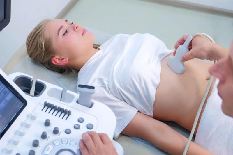
Our abdomen Ultrasound Scan
At Universal Ultrasound Diagnostics, we are dedicated to providing exceptional private abdomen ultrasound scans at our abdomen ultrasound clinic in Birmingham, UK. We offer flexible and convenient dates and times that suit your needs and preferences. With our cutting-edge Ultrasound systems and highly skilled, qualified and experienced staff, we deliver precise and reliable imaging for both our private clients and referring practitioners in a comfortable and discreet setting. Our commitment to utilising the latest technology ensures we consistently deliver the highest quality performance you can trust.
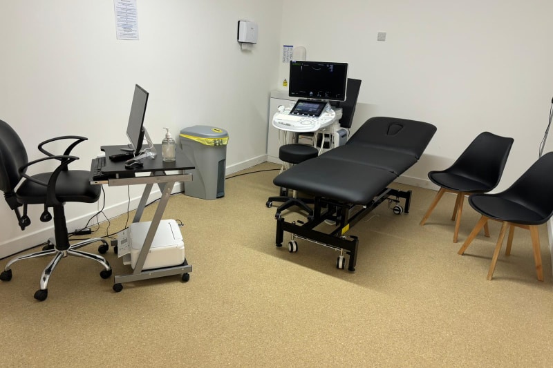
Abdominal Scan Time & Cost
Scan Time
An abdominal ultrasound scan may take up to 30 minutes, depending on the circumstances.
Results
The sonographer will discuss any immediate findings with you, and the report and images will be emailed to you on the same day.
Typical Cost
You can see our latest rates on our booking page.
Paediatric Abdominal Ultrasound Near Soilhull & How They Help
Tummy aches, vomiting, low appetite, and a swollen or bloated abdomen are common in children. Your child’s doctor may recommend an ultrasound of the abdomen to get a better look inside the stomach and rule out anything more serious, even though the cause may be straightforward, such as a viral illness or constipation.
A paediatric abdominal ultrasound near Solihull uses high-frequency sound waves (not X-rays) to produce detailed images of the internal organs.
It can help to:
- Examine the kidneys, liver, gallbladder, pancreas, spleen, aorta, abdominal wall, check for hernia or appendix which are all located inside the child’s upper abdomen area.
- Investigate persistent or recurrent bloating, vomiting, or abdominal pain.
- Identify inflammation, infection, gallstones, or other kidney-related problems.
- Detect fluid, cysts, tumours, or organ enlargement.
- Monitor known disorders affecting the liver or kidneys
It is commonly used as a first-line test in children’s hospitals across the United Kingdom because it is safe, painless, and radiation-free.

During the examination, your child lies on a couch and a small amount of gel is applied to their abdomen. The sonographer gently moves a probe across the abdomen to capture images. Most scans take 20 to 30 minutes, and you can stay with your child throughout the examination.
As is customary in many NHS hospitals, some children may be asked to refrain from eating or drinking for a few hours before the scan to improve image quality.
Additional Resources:
We provide quick, child-friendly private abdominal ultrasounds at Universal Ultrasound Diagnostics, frequently on the same day. Our skilled sonographers explain the process before scanning and explain findings in simple terms, helping you receive fast answers and peace of mind. A report and images are emailed to you on the same day and we advise you share them with your GP or paediatric specialist, who can recommend further testing or put a treatment plan in place.
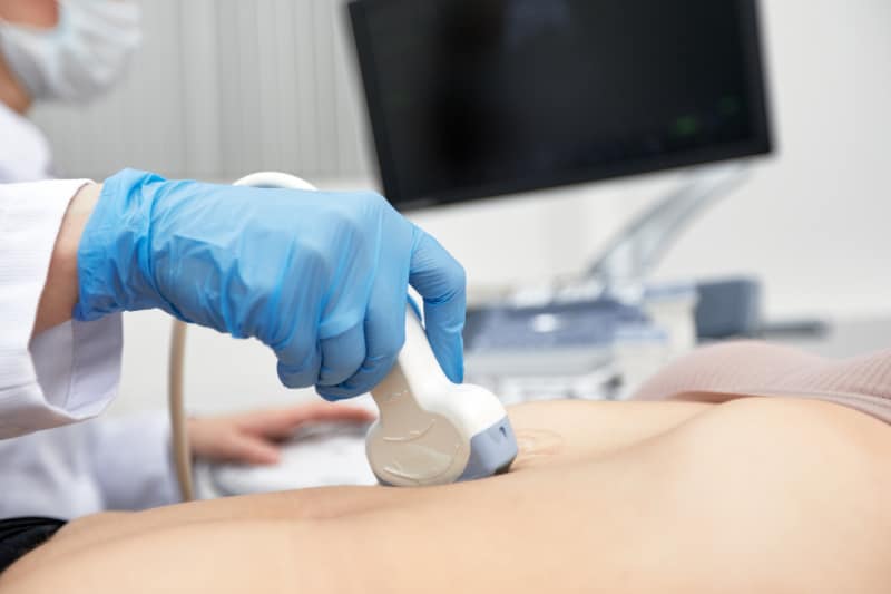
Abdominal Scans With Same Day Results And Peace Of Mind
Our same day abdominal scans are performed by qualified, experienced, caring, respectful and compassionate staff with experience in the NHS and private clinics. An abdominal ultrasound produces painless, real-time images of your abdominal organs such as kidneys, spleen, pancreas, liver, gallbladder, and associated blood arteries. The ultrasound is non-invasive and is performed in a clean, professional and comfortable environment. It does not involve any radiation or needles. The scan is usually completed within 30 minutes. Results are discussed immediately after the scan. We recommend follow-up care with your healthcare provider. We provide you with timely answers, which reduce stress or anxiety and enable you to decide on your next course of action.
The abdominal scan helps to detect:
- Cause of abdominal pain or bloating. It is advisable for women to have a pelvic scan alongside an abdominal scan if they experience bloating as they may have issues with their reproductive organs
- Kidney stones or gallstones
- Liver diseases, including cirrhosis and fatty liver
- Pancreatitis
- Anomalies or growths across the abdomen
- Abdominal masses or cysts
- Hernia
- Appendicitis
Abdominal Wall, Hernia and Appendix Ultrasound Scans near Solihull
Sometimes the cause of abdominal pain or a visible “bulge” sits closer to the surface than you might expect. That’s why we can also focus an abdominal ultrasound on the abdominal wall, check for a hernia, or take a closer look at the appendix when it’s clinically appropriate.
When a patient has palpable lumps, localised soreness, swelling, or pain that gets worse following breathing, coughing, or physical exercise, an abdominal ultrasound can be useful. It makes it possible to assess the muscles and soft tissues of the abdominal wall for anomalies in the soft tissues, such as fluid accumulations, inflammation, or other conditions.
A hernia ultrasound can help confirm whether tissue is pushing through the abdominal wall, particularly if the swelling comes and goes or is worse when standing, lifting, or straining. Dynamic scanning (checking the area during gentle movement or bracing) can be especially helpful when a hernia is suspected but not always obvious.

An appendix ultrasound near Solihull may be used when symptoms suggest appendicitis, such as pain that becomes more focused in the lower right side of the abdomen, tenderness, fever, nausea, or reduced appetite. We will suggest the best course of action, including an immediate medical evaluation if required.
Our team can guide you on the best ultrasound scan based on your symptoms if you are not sure what type of scan you need.
Meet the Team
Lydia, our CQC Registered Manager and Operations Director, leads the team with empathy, faith and the utmost integrity. Her patient-focused approach helps to shape the culture of our service.
Our Consultant Radiologist Dr Moti, is supported by our advanced sonographer Andrew, combine clinical excellence with reassurance, giving our patients peace of mind and genuine warmth. Ali brings clarity to every scan and calm assurance, helping patients feel at ease, supported, and well-informed.
Behind the scenes, Emma, our practice manager, plays a pivotal role, ensuring every detail of the patients’ journey is seamless. She works alongside the Registered Manager to ensure quality assurance is maintained.
Carly, our Medical Administrative Officer, plays a crucial role in patient experience and works alongside the Registered Manager to maintain high-quality standards through promoting continuous improvement across the practice.
Together as a team, we are united by one purpose:
Our Mission - To deliver high-quality care with compassion that not only meets the patients’ expectations but goes over and beyond to exceed expectations.

Why Choose Our Private Ultrasound Scan Clinic?
1. Expert Sonographers: Our clinic boasts highly skilled and experienced sonographers specialising in Diagnostics and Pregnancy scans.
2. State-of-the-Art Technology: We use cutting-edge ultrasound systems for precise and reliable imaging.
3. Comfortable and Discreet Setting: Our clinic ensures a comfortable and confidential patient environment.
4. Flexible Appointments: We offer flexible dates and times to suit your convenience.
5. Prompt Results: Immediate findings are discussed during your appointment, and a detailed report and images are emailed to you the same day.
Our Mission, Vision And Values
Mission - To be the most efficient and preferred diagnostic imaging centre that provides excellent quality care, that not only meets but goes above and beyond to exceed patients’ expectations.
Vision - Our vision is innovation as healthcare and technology are continuously evolving, which will enhance scanning techniques, image quality, accuracy, and efficiency and provide clearer insights into diseases.
We are proud to be a compassionate, caring, and respectful team. We do this by asking staff to work within our core values:

Compassionate by being inclusive, attentive, caring, respectful and showing kindness to staff, our patients, and their families.

Caring by going beyond compliance, performing above and beyond to improve our services and providing person-centred care by involving patients in their care, offering choices, and promoting dignity and privacy.

Respectful by active listening, seeking to understand others, showing empathy for differences, apologising when things go wrong, being polite and showing gratitude to our staff and patients.
About your abdominal scan appointment
PREPARATION
You must starve for 6 hours prior to your appointment. You may drink water or other non-fizzy drinks, black tea or coffee, or fruit juice. Please DO NOT drink milk, any drinks which contain milk or fizzy drinks as this may affect the images. If you are diabetic, we advise you to have the earliest appointment if possible. Do not follow these instructions if they conflict with your doctor’s orders. Please take your regular medications as normal. If you are unsure, please contact us on 0121 392 3112 or your doctor for further advice.
ARRIVING FOR YOUR ABDOMINAL SCAN APPOINTMENT
Please arrive 5 to 10 minutes before your actual appointment time as it may not be possible to scan you if you are late, resulting in your payment being forfeited as per our payment and refund policy. CHILDREN ARE NOT PERMITTED IN THE ROOM.
In the examination room, you will be asked to lie on a couch. The sonographer performing the scan will ask you to expose your upper abdomen. The gel will be applied over your upper abdomen, and it may feel cold. The sonographer will gently move an ultrasound probe over your abdomen to obtain images. The images will come up on a big-screen television in front of you so that you can see all the structures that are being examined. Unless the area being examined is tender, the examination is not painful.
The sonographer will always ensure your lower body will be covered with a sheet to ensure your dignity. A chaperone will always be available in the scan room. Your decision/choice not to have a chaperone will be respected. The scan may take up to 20 minutes, depending on individual circumstances.
AFTER THE EXAMINATION
The report will be sent to the doctor who referred you for the scan on the same day. If you referred yourself, the report will be issued immediately following the examination. If we refer you straight to outpatients, you will be able to take your report with you.
Abdominal Ultrasound Scan Faqs
What is an abdominal scan?
An abdominal ultrasound scan is a type of scan that looks at the various organs of the abdomen. It uses high-frequency sound waves to produce pictures of the structures within the upper abdomen. The scan does not use radiation and has no known harmful effects. An ultrasound scan is safe, non-invasive and does not use ionising radiation.
An abdominal ultrasound imaging is performed to assess the:
- Liver
- Gallbladder
- Spleen
- Bile Ducts
- Pancreas
- Kidneys
- The Abdominal Aorta and other blood vessels of the abdomen
An abdominal ultrasound is used to help diagnose a variety of conditions, such as:
- An Abdominal Aortic Aneurysm
- Abnormal Liver Function
- Abdominal Pain or Distention (enlargement)
- Enlarged Abdominal Organ
- Kidney Stones
- Gallstones
What are the symptoms of abdominal issues?
Some of the signs of abdominal problems or disorders are:
- Pain or discomfort in the abdomen
- Loss of appetite and weight loss
- Itchy skin
- Bloating
- Tenderness
- Abdominal mass or bulge
- Nausea or vomiting
- Constipation
- Bloody or black tarry stools
- Painful or lack of urination
Schedule an Abdominal Ultrasound Scan at our Clinic near Solihull
What are the potential causes of abdominal issues?
- Tumour
- Cyst
- Infection (Recurring urinary tract infection (UTI)
- Reduced/increased blood flow
- Blockages
- Narrowing of vessels
- Enlarged organs
Possible causes in children may include:
- Constipation
- Abdominal migraines – recurrent episodes of abdominal pain with no identifiable cause
What is a tumor?
A tumour is a solid mass of tissue caused by a collection of abnormal cells in one area. Tumours can be both benign and malignant. An ultrasound will be able to detect the presence of a tumour however it will not be able to differentiate between the types.
What is a cyst?
A cyst is an abnormal pocket of fluid forming on an organ such as the Thyroid. They can range in size from just a few millimetres to a heavy “bag” noticeable even through the skin. Cysts are usually harmless; however, some may require medical intervention to remove them.
If you have any concerns, please schedule an Abdominal Ultrasound Scan at our Clinic near Solihull, Birmingham
What is an infection?
An infection is caused by germs entering the body and causing an immunological reaction. These reactions can include swelling and fever and may require medical intervention to resolve.
What is a blockage?
A blockage is caused by something blocking either blood flow or an organ. This could be due to clots in the blood vessels, or stones in the gallbladder or kidneys.
What is the narrowing of a blood vessels in the abdomen?
A narrowing of the blood vessels, known as vascular stenosis, may be seen on an abdominal ultrasound. Although vascular stenosis can occur in any abdominal blood channel, it is more common in the renal arteries, or kidneys.
Vascular stenosis can have several causes, however what follows represent a few of the most prevalent ones:
Atherosclerosis - This condition is brought on by the buildup of fatty deposits, or plaques, on the innermost layer of the arteries. Over time, these deposits can harden and constrict. While it can affect any artery in the body, it most commonly affects the renal arteries. Anaemia, hypertension, hypercholesterolemia, diabetes, obesity, smoking, and a family history of cardiovascular disease are the main risk factors.
Fibromuscular dysplasia - This condition is characterised by abnormal growth of muscle and fibrous tissue, which narrows and unevenly distributes the arterial walls. Any artery in the body may be affected by fibromuscular dysplasia, although the renal arteries are most frequently affected, particularly in young women. Vascular obstruction, scarring, or constriction can result from damage and inflammation to blood vessels caused by a group of illnesses called vasculitis.
Vasculitis - While vasculitis can affect any blood vessel in the body, several types of the disease, such as Takayasu’s arteritis, giant cell arteritis, and polyarteritis nodosa, can affect the abdominal arteries.
Compression - Some causes of compression include tumours, cysts, larger organs, and scar tissue pressing on blood vessels, leading them to narrow. The liver disease. Veins, which drain blood through the liver, and the sigmoid blood vessels, which drained blood from the intestines, are more likely to experience compression.
Thrombosis - Although it can affect any blood artery in the body, veins were more likely than arteries to have this illness. When a thrombus restricts or obstructs blood flow within a blood vessel, this occurs.
What are enlarged organs?
Organs can become enlarged due to inflammation from infection or cyst/tumour growth. It is also possible however that they become enlarged due to digestion issues, or very rarely due to internal bleeding.
What do I do if I have sudden severe abdominal pain?
A sudden excruciating or unbearable pain in a particular area of your abdomen may indicate a severe problem that could quickly worsen without treatment.
If there is any possibility that you could be pregnant, take a pregnancy test.
Go to Accident and Emergency or call 111/999 for emergency help if:
- You have a sudden onset of severe stomach ache.
- It hurts, or you feel pain when you touch your stomach.
- You are vomiting blood, or the vomit looks like ground coffee.
- Your stool (poo) has blood or is black and sticky and extremely smelly.
- You are less than 16 weeks pregnant and have severe abdominal pain.
- You cannot pass urine (pee) or stool (poo).
- You cannot breathe or have severe chest pains.
- You are diabetic, and you are vomiting.
What are the blood tests relating to abdominal pain?
Commonly requested abdominal pain tests from blood samples include:
- Full Blood Count (FBC) or Complete Blood Count (CBC)
- Electrolytes and Urea (E&U; DAX) or Blood Urea Nitrogen (BUN)
- Creatine
- C-reactive protein (CRP)
- Erythrocyte Sedimentation Rate (ESR)
- Serum Amylase
- Calcium levels
- Lipase
- Lactate Dehydrogenase
- Liver Function Test
- Blood sugar level
- Uric acid levels
- Blood gas (arterial blood gas or ABG)
- HBA1c
- Thyroid Function Test (TSH, T3, T4)
- TTG
- Ceruloplasmin level
- Serum Copper level
- Transferrin saturations
- Ferritin and Iron levels
- Anti LKM
- Anti Smooth Muscle Antibody (ASMA)
- Hepatitis serology or hepatitis screening
- Alpha Feto Protein
- Cryo Embryonic Antigen (CEA) level
- Ca 125
- Ca19-9
- Anti Mitochondrial Antibody
- Alpha1 Antitrypsin level
- Female hormone level (LH, FSH)
- Carcinoembryonic Antigen (CEA) level
- Serum Cortisol level
- Leaky Gut (Intestinal permeability) testing, also called the mannitol and
lactulose test.
Our website provides a wide range of blood tests associated with abdominal issues. If you are unsure which tests you require, we highly recommend you consult your doctor (GP).
What kind of things can an abdominal ultrasound detect?
An abdominal ultrasound scan can detect tumours, cysts, infections, enlarged organs and narrowing of blood vessels. It can also be used to determine the source of abdominal pain such as gallstones or kidney stones, abscesses or inflamed appendix, and help determine the cause of vomiting in young infants. However, ultrasound scans alone cannot be used to diagnose disease and must be used with other diagnostic tests.
What cancers can abdominal scan detect?
Ultrasound is often one of the first-line tests in cancer detection. An ultrasound can help diagnose stomach cancer. It can detect abnormal tissues, growths, and cysts and diagnose cancer based on how those images look. While it cannot diagnose cancer, it can detect abnormal tissues that may be cancerous.
It can show some signs of cancer, such as tumours or inflammation in certain parts of the pancreas, gallbladder, liver, kidneys or other abdominal structures. However, it cannot detect tumours of the colon or other parts of the pancreas. The scan helps to look for tumours in some abdominal regions that do not appear well on X-rays. Ultrasound images can also help to determine if a tumour is solid or filled with fluid, how deep it is, and whether it has spread to the stomach walls or other organs.
However, ultrasounds are not always accurate in detecting stomach cancer and may need to be combined with other tests, such as biopsy or endoscopy, to confirm a diagnosis.
For an Abdominal Ultrasound Scan at our Clinic near Solihull, click here
Can an abdominal ultrasound detect inflammation?
Ultrasound will not show inflammation itself, but rather the signs of it such as free fluid, enlarged lymph nodes and changes in the fat surrounding the organs.
Can an abdominal ultrasound detect an infection?
Infection can be detected in certain organs, especially if there are signs of inflammation.
Can an abdominal ultrasound detect IBS?
Due to gas in the colon, it obstructs the ultrasonic frequencies, rendering ultrasonography unsuitable for diagnosing conditions such as IBS. Several other factors mean an abdominal ultrasound cannot detect IBS these include:
Ultrasound is not used as a diagnostic tool for irritable bowel syndrome (IBS) for several reasons:
1. Lack of Structural Abnormalities: IBS is a functional gastrointestinal disorder, indicating the absence of any readily apparent changes or structural abnormalities in the digestive tract that are detectable by imaging. Chronic Irritable Bowel Syndrome (IBS) is characterised by changes in bowel habits, abdominal discomfort, and pain often triggered by stress, nutrition, hormone changes, or exposure to toxins.
2. Diagnosis Based on Symptoms: The Rome criteria are the main diagnostic tool for IBS. Abdominal pain or discomfort related to altered bowel habits is included in this criterion, and it must have persisted for at least three days each month for the previous three months.
3. Limited Utility: An abdominal ultrasound is better at identifying structural anomalies such as gallstones, tumours, or gastrointestinal tract inflammation. Ultrasonography is not a reliable method of confirming or ruling out IBS because this condition is not linked to structural problems.
4. Misdiagnosis Potential: IBS may be misdiagnosed if ultrasonography is the only method used to diagnose it since it may not offer useful information about the illness. This may lead to unneeded medical treatments or procedures. To diagnose IBS without an ultrasound, medical professionals usually combine a patient’s medical history, physical examination, and symptom assessment. In order to rule out illnesses other than IBS that can mimic its symptoms, further testing is occasionally advised. These could involve stool or blood tests, and in some cases, endoscopies or colonoscopies to examine the digestive tract.
To diagnose IBS, your doctor will usually utilise a mix of physical examination, medical history, and symptom evaluation rather than an ultrasound. To rule out other illnesses that might mirror the symptoms of IBS, further testing might occasionally be prescribed. Blood tests, stool tests, and, in some cases, colonoscopies or endoscopies to examine the gastrointestinal tract directly are examples of these.
What does the ultrasound equipment look like?
The ultrasound machine consists of a computer console, video monitor and an attached probe called a transducer. The probe uses high-frequency sound waves to build a picture of the inside of the body.
What do I wear?
You will need to loose and comfortable clothing. You will need to expose the whole of your abdomen for the scan, so you will need to pull your trousers down to your bikini line and your top up towards your chest.
How does the procedure work?
The high frequency sound waves “bounce” back of organs and tissues creating an echo effect that the transducer than translates into the image seen on the screen. The pitch, direction and time it takes the waves to reach the probe all help to determine what the picture should look like.
What are the red and blue blobs on the ultrasound?
During the scan, the sonographer may use a colour doppler to check blood flow in your tissue and organs. The colours will appear as blue and red blobs on the image and are a display of real-time sound waves detected in the blood vessels. They are nothing to be concerned about.
How is the procedure performed?
You will be asked to lie face up on the exam couch and expose your abdomen. This is the area from your bikini line to just under your chest. A gel will be applied to the skin, and the sonographer will move a probe through it and across your abdomen. During the scan, you may be asked to turn onto your side so the sonographer can examine your organs when lying differently.
During the exam, the sonographer may go quiet or examine the same area multiple times. This is usually nothing to be concerned about as the sonographer must concentrate and ensure they see all parts of each organ. If there are any concerning findings, the sonographer will talk you through what they see and give you recommendations for follow-up.
What will my child experience during and after the procedure?
Your child will be asked to lie face up on the exam couch and gel applied to their skin. The scan itself should be painless (unless there are any tender areas) and will be quick and easily tolerated. During the procedure, the sonographer may need to check blood flow with the colour doppler. While this is in use, they may hear pulse-like sounds but should not be alarmed.
The sonographer will warn them if there are going to be any noises. Once the scan is complete, the gel will be wiped off your child’s skin without lasting effects. The child will then be able to resume their normal activities. Any findings on the scan will be discussed with you directly.
Get in touch for an Abdominal Ultrasound Scan at our Clinic near Solihull
Do I need to consent to an abdominal scan?
All patients must give their consent (permission) before undergoing an examination. If they cannot give consent themselves, then a person with power of attorney will need to give consent on their behalf. Please ensure the pre-treatment consent form is completed via the embedded link in your confirmation email before you attend the appointment. The scan will not go ahead if this form is not completed.
Are abdominal scans painful?
Abdominal scans are not painful, but they may be uncomfortable as the sonographer will have to press the ultrasound scan probe to your abdomen gently. If the area is tender, this may cause some discomfort.
What happens during the scan?
The sonographer will put ultrasound gel onto your skin and move a probe over your abdomen. They will examine your whole front to check all the organs and will then also ask you to turn onto each side to check liver, kidneys and pancreas from other angles.
What are the limitations of an abdominal ultrasound?
Ultrasound waves are disrupted by air and gas, and therefore ultrasound scans are not ideal for imaging air-filled bowel, or organs that may be obstructed by the bowel. Ultrasound cannot penetrate bone either and therefore cannot be used to diagnose fractures or breaks.
Larger patients may also be more difficult to scan using ultrasound because of the more significant amounts of tissue. This can weaken the sound waves and thus affect the images.
Will an ultrasound show a bowel blockage?
Bowel blockage, or bowel obstruction, occurs when a partial or complete blockage in the large or small intestine prevents the normal passage of food, gas, and fluids. Here are some key points about bowel blockage/obstruction:
Symptoms:
- Loss of appetite
- Vomiting
- Constipation
- Intermittent crampy abdominal pain.
- Abdominal swelling
- Inability to pass gas or have a bowel movement
Causes:
- Colon cancer
- Crohn’s disease: an inflammatory bowel disease
- Intestinal adhesions: Bands of fibrous tissue that form after abdominal or pelvic surgery.
- Diverticulitis: an inflammation of small pouches in the digestive tract
- Hernias: Portions of the intestine protruding into other body parts
- Volvulus: Twisting of the colon.
- Intestinal pseudo-obstruction (paralytic ileus): Disruption of normal muscle contractions in the intestines without a physical blockage.
- Impacted faeces
Treatment:
- For complete blockages, you may be hospitalised to receive treatment
- In severe cases, surgery may be necessary
- See urgent medical care to prevent any complications
An ultrasound can detect a bowel blockage and is often the preferred imaging method for diagnosing intestinal obstructions in children. In cases of intussusception, where the intestine folds into itself, an ultrasound usually reveals a distinctive "bull's-eye" pattern indicating the coiled intestine. While ultrasound is effective for detecting small bowel obstructions, computed tomography (CT) scans offer more detailed imaging and are better at identifying the cause of the obstruction. If a bowel blockage is suspected, seeking medical evaluation is crucial for accurate diagnosis and appropriate treatment.
It's easy to book an appointment.
Universal Ultrasound Diagnostics offers an online booking system for easy appointment scheduling. There are options for walk-ins depending on availability: urgent scans within 5 days and routine scans within 14 days.
Appointment notifications and reminders are sent via email. The system supports self-check-in and online referrals by GPs.
We provide chaperones and interpreting services and allow family presence during pregnancy scans.
Our facility is accessible by public transport and has parking.
We allow family presence for most scans; however, children are not allowed in the room for certain scans. Please check your appointment confirmation email for clarity on our safety policy.
Our FAQ page answers a lot of general questions about your scan.
There is parking around The Green or Pay & Display at the side of The Bulls Head Pub, which charges £1 per hour (cash and card accepted). We are open 8 am to 11 pm Monday to Sunday.
Opening Times
Monday to Sunday 8 am - 11 pm
Location
10c The Green, Kings Norton, Birmingham, B38 8SD