

Pelvic Ultrasound Scan Bromsgrove
Private Pelvic Ultrasound Scans at our Birmingham Clinic near Bromsgrove.
Pelvic Ultrasound Scan Near Bromsgrove
A Pelvic Ultrasound Scan near Bromsgrove is an examination that examines the pelvis. The reasons for performing the scan include, but are not limited to:
- Pelvic pain
- Irregular vaginal and postmenopausal bleeding
- Heavy periods
- Bloating
- Fertility problems/concerns
- To check endometrial thickness
- Fibroids
- Check for polycystic ovaries (PCOS)
- To check for endometriosis or adenomyosis
- Cause of pregnancy loss (recurrent miscarriages)
- Ovarian cysts or tumours and screening for ovarian cancer
- Vaginal anomalies
- Check the position of the coil
- Congenital uterine anomalies
- Check pregnancy-related problems (foetal development monitoring,
ectopic pregnancy, and pregnancy confirmation) - Check for suspected retained products of conception (RPOC) after a miscarriage
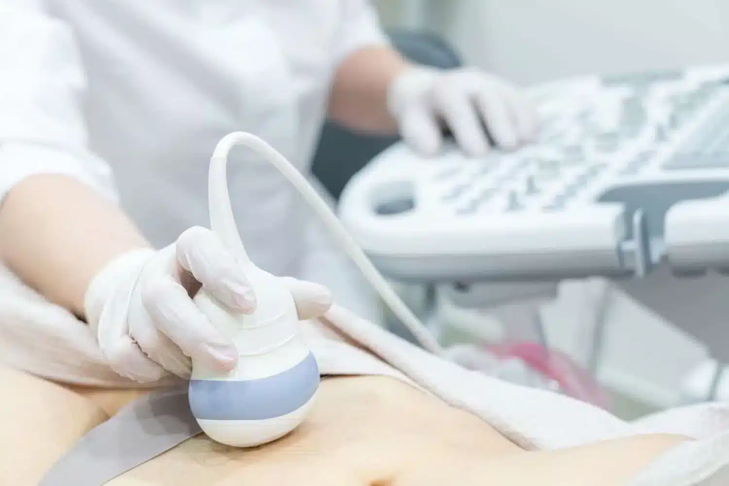
The scan is generally carried out in two parts. The first part is a transabdominal scan which needs a full bladder. The second part is a transvaginal scan which is performed after you have emptied your bladder. If you have never been sexually active, a transvaginal scan will not be performed. Please notify the sonographer performing the scan. You do not need a GP referral for this examination.
SAME-DAY FEMALE AND MALE PELVIC SCAN FOR YOUR PEACE OF MIND
Your health matters, and you should not have to sit on long waiting lists for a scan. Our Same-Day Pelvic Scan service will provide you with fast, reliable results, so you can get the answers you need without the long waiting times. We offer a comfortable, private environment and the peace of mind you deserve.
Both men and women can benefit from pelvic ultrasound, although women are more likely to have it done because of its usefulness in evaluating female reproductive health. The male pelvic scan can aid in the diagnosis of various diseases and conditions, such as bladder problems, prostate cancer and testicular issues. The pelvic scan is frequently carried out to assess the tubes, ovaries, uterus, endometrium thickness, and bladder in females.
Pelvic Scan Timeline & Cost
Scan Time
Pelvic Adult (Transabdominal Only) 30 Mins
Pelvic Adult (Transabdominal and Transvaginal) 40 Mins
Pelvic and Kidney Female Scan 50 mins
Pelvic – Paediatric Female 40 Mins
An ultrasound scan alone cannot be used to diagnose disease or cancer and must be used in conjunction with other diagnostic tests.
Typical Cost
You can see our latest rates on our booking page.
Results
The sonographer will discuss any immediate findings with you, and the report and images will be emailed to you on the same day.
Our Pelvic Ultrasound Scan
At Universal Ultrasound Diagnostics, we are dedicated to providing exceptional private pelvic ultrasound scans at our clinic in Kings Norton, UK.
We offer flexible and convenient dates and times that suit your needs and preferences. With our cutting-edge Ultrasound systems and highly skilled, qualified, and experienced staff, we deliver precise and reliable imaging for both our private clients and referring practitioners in a comfortable and discreet setting.
Our commitment to utilising the latest technology ensures we consistently deliver the highest quality performance you can trust.
What is a Pelvic CA125 Ultrasound Scan?
A pelvic CA125 (Cancer Antigen 125) ultrasound scan near Bromsgrove is usually performed when ovarian cancer is suspected, following a CA125 blood test which showed raised CA125 levels (>35 IU/ml). The scan utilises high-frequency sound waves to create detailed images of the ovaries, cervix, the uterus, bladder, and surrounding structures. It helps to detect polyps, cysts, fibroids, masses or other changes or abnormalities in the pelvic region.
CA125 may be elevated due to non-gynaecological and gynaecological causes. The various conditions include:
Pathological
Benign causes
- Gynaecological - endometriosis, uterine fibroids, benign ovarian tumours, pelvic inflammatory disease.
- Non-gynaecological – renal failure, tuberculosis, heart failure, cirrhosis with or without ascites, active or chronic hepatitis, pericardial disease, chronic or acute pancreatitis.
Malignancy causes
- Cancers such as endometrial, ovarian, lung, pancreatic, bowel, breast, and lymphoma.
According to the NICE guidelines, CA125 testing is recommended for women who present with one of the following symptoms on a frequent or persistent basis
- loss of appetite
- Swelling or bloating - persistent abdominal distension
- Changes in menstrual cycle
- Persistent abdominal and pelvic pain
- Increased urinary urgency
- Unexplained - fatigue , change of bowel habit or weight loss

If the scan suggests ascites and or an abdominal or pelvic mass (which is not uterine fibroids), an urgent referral for review in gynaecology is recommended.
A pelvic CA125 ultrasound alone does not diagnose cancer. The scan helps to detect any abnormalities early, and this will guide the next steps in your care, such as further investigations to confirm the diagnosis.

Why Choose Our Private Ultrasound Scan Clinic?
1. Expert Sonographers: Our clinic boasts highly skilled and experienced sonographers specialising in Diagnostics and Pregnancy scans.
2. State-of-the-Art Technology: We use cutting-edge ultrasound systems for precise and reliable imaging.
3. Comfortable and Discreet Setting: Our clinic ensures a comfortable and confidential patient environment.
4. Flexible Appointments: We offer flexible dates and times to suit your convenience.
5. Prompt Results: Immediate findings are discussed during your appointment, and a detailed report and images are emailed to you the same day.
Meet the Team
Lydia, our CQC Registered Manager and Operations Director, leads the team with empathy, faith and the utmost integrity. Her patient-focused approach helps to shape the culture of our service.
Our Consultant Radiologist Dr Moti, is supported by our advanced sonographer Andrew, combine clinical excellence with reassurance, giving our patients peace of mind and genuine warmth. Ali brings clarity to every scan and calm assurance, helping patients feel at ease, supported, and well-informed.
Behind the scenes, Emma, our practice manager, plays a pivotal role, ensuring every detail of the patients’ journey is seamless. She works alongside the Registered Manager to ensure quality assurance is maintained.
Carly, our Medical Administrative Officer, plays a crucial role in patient experience and works alongside the Registered Manager to maintain high-quality standards through promoting continuous improvement across the practice.
Together as a team, we are united by one purpose:
Our Mission - To deliver high-quality care with compassion that not only meets the patients’ expectations but goes over and beyond to exceed expectations.
TRUSTED BY MANY
About your Pelvic Ultrasound Scan appointment
PREPARATION
Your bladder must be full when you attend. One to two pints of clear fluid should be consumed an hour before the appointment. You can have fruit juice, coffee or black tea, or water or other non-fizzy beverages. You will feel the urge to urinate once your bladder is properly filled. It is too full if it starts to hurt, and if you can, you can partially empty it.
If you are unsure, please contact us on 0121 392 3112 or your doctor for further advice.
ARRIVAL
Please arrive 5 to 10 minutes before your actual appointment time as it may not be possible to scan you if you are late, resulting in your payment being forfeited as per our payment and refund policy. As this is an intimate examination.
CHILDREN ARE NOT PERMITTED IN THE EXAM ROOM.
The examination
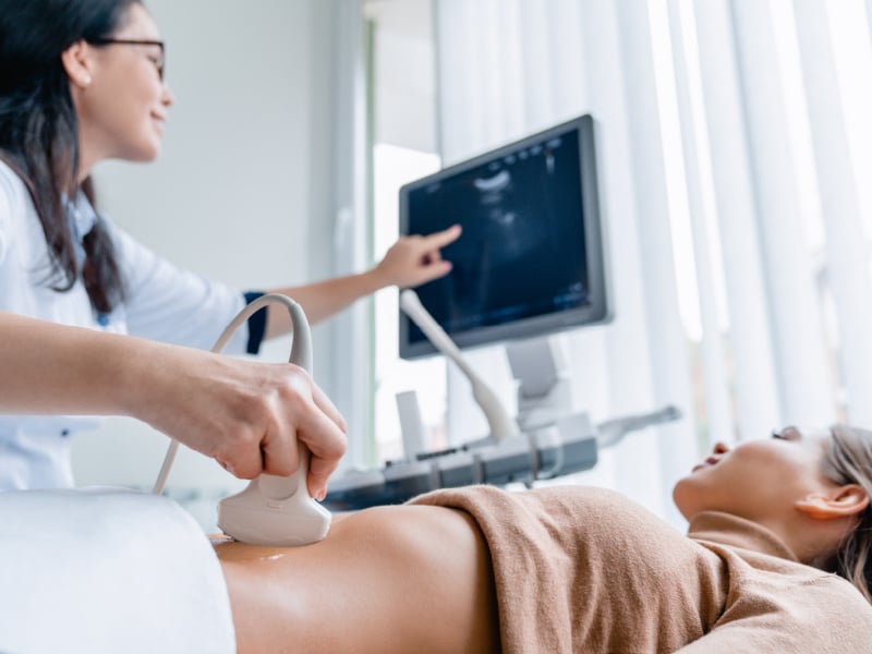
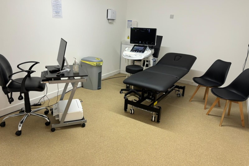
In the exam room, you will be instructed to recline on a couch. During the scan, the sonographer will ask you to reveal your lower abdomen. You’re abdomen will be covered with the gel, which could feel cold. To get pictures of your pelvis, the sonographer will gently slide an ultrasound probe across your lower abdomen. The examination is not uncomfortable unless there is tenderness in the area being probed.
With your permission, a transvaginal scan will be carried out to get a clearer view of your uterus and ovaries. Even when you’re on your period, you can still do this. Following the transabdominal scan, you will be instructed to return to the ultrasound examination room after emptying your bladder.
You’ll be instructed to undress and lie down on a couch with a wedge cushion on top. To protect your dignity, the sonographer will always make sure a sheet is placed over your lower body. The scan room will always have a chaperone on hand. We will respect your decision to forego a chaperone. An ultrasonic probe will be lubricated with gel and protected by a disposable latex or non-latex sheath. Kindly inform the sonographer if you have a latex allergy. After that, the probe will be inserted into the vagina to capture pictures of the pelvis, specifically the uterus and ovaries. The test won’t hurt, but it can be uncomfortable.
Transvaginal exams are not performed on children or individuals who have never engaged in sexual activity. You can talk to the sonographer when you arrive for your scan if you would prefer not to have an internal examination done. Your choice or decision will be honored. Nevertheless, the quality of photographs acquired without conducting a transvaginal examination will be limited. Depending on the specific situation, both sections of the test could take up to 20 minutes.
AFTER THE EXAMINATION
The sonographer will discuss any immediate findings with you, and the report, along with images, will be emailed to you on the same day.
It's easy to book an appointment.
Universal Ultrasound Diagnostics offers an online booking system for easy appointment scheduling. There are options for walk-ins depending on availability: urgent scans within 5 days and routine scans within 14 days.
Appointment notifications and reminders are sent via email. The system supports self-check-in and online referrals by GPs.
We provide chaperones and interpreting services and allow family presence during pregnancy scans.
Our facility is accessible by public transport and has parking.
We allow family presence for most scans; however, children are not allowed in the room for certain scans. Please check your appointment confirmation email for clarity on our safety policy.
Our FAQ page answers a lot of general questions about your scan.
There is parking around The Green or Pay & Display at the side of The Bulls Head Pub, which charges £1 per hour (cash and card accepted). We are open 8 am to 11 pm Monday to Sunday.
Opening Times
Monday to Sunday 8 am - 11 pm
Location
10c The Green, Kings Norton, Birmingham, B38 8SD

Pelvic Ultrasound Scan Faqs
What is a Pelvic Ultrasound scan?
A pelvic ultrasound scan is an examination that utilises high-frequency soundwaves to create images of the inside of your body such as of the ovaries.
Is a Pelvic Ultrasound Scan safe??
A pelvic ultrasound scan is safe and does not have any of the risks associated with ionising radiation. It has been used to provide valuable images and other diagnostic information with no evidence of any harm. However, as a precaution, the sonographer will take every care to ensure that the level and duration of the ultrasound examination are not more than necessary to obtain the diagnostic images or information to minimise any potential risk to the patient.
Schedule a Pelvic Ultrasound Scan at our Clinic near Bromsgrove
Why Have I Been Referred for A Pelvic Ultrasound?
A pelvic ultrasound is the common initial modality for imaging gynaecological pathologies.
What particular reason led to my referral?
A referral can be based on a physical examination, your symptoms, or family history. Discuss your unique circumstances with your healthcare provider for a tailored explanation.
COMMON REASONS FOR REFERRALS
Problems with menstruation such as heavy, painful, irregular, or missed periods and postmenopausal bleeding. The scan can help to determine why menstruation has been unpredictable or unduly severe or why you are bleeding past menopause.
Pelvic Pain: The pelvic ultrasound can detect an infection such as pelvic inflammatory disease (PID), adnexal masses, abscesses, adenomyosis, fibroids or ovarian cysts.
Persistent bloating or raised CA125 : This could be a sign of ovarian cancer. It is advisable for you to have both pelvic and abdominal ultrasound scans to rule out any abnormalities that could be the cause of bloating, without having the need for another referral or to book the scans separately.
Other symptoms that may need to be investigated with the help of both pelvic and abdominal ultrasound scans are loss of appetite or feeling full quickly, abdominal or pelvic pain that is from the top of your belly to the top of your thighs, potential reasons for urinary symptoms, such as trouble emptying the bladder or frequent urination, the scan can be used to rule out masses or pressure from pelvic tissues.
Fertility Problems: The scan can help to evaluate ovarian or uterine anomalies that may impact pregnancy or cause pregnancy loss or recurrent miscarriages. It can also be used as a baseline for assisted conception such as IVF.
Concerns associated with pregnancy: The scan can help to confirm pregnancy or monitor fetal development in the early stages of pregnancy, or check for ectopic pregnancy.
Examining for issues such as ovarian cysts or tumors and screening for ovarian cancer The scan will help to determine if a tumor is benign or malignant by assessing the size, shape and texture.
Endometrial Conditions: The pelvic ultrasound does not check for endometriosis in the endometrium, it checks for adhesions, polyps, fibroids into endometrium, endometrial lining to measure the thickness, and cervical niches.
Cancer screening: If there are risk factors or troubling signs, for instance, ovarian or uterine cancer. When performed transvaginally, osteoporosis screening looks at the endometrium to indirectly measure estrogen levels. Keeping an eye on known conditions, your doctor may wish to track the course of any diseases you have been diagnosed with, such as fibroids, endometriosis, or PCOS (Polycystic Ovary Syndrome).
A Pelvic Ultrasound is usually performed in 2 parts:
Transabdominal (TA), is performed to provide a general overview of the pelvis and maybe necessary in certain situations (paediatric patients) for imaging the entire pelvic pathologies without performing a transvaginal ultrasound. A full bladder is required as it is helpful as an acoustic window to enable better imaging of the adnexa and uterus.
Transvaginal (TV) provides a deeper look at the uterus, the ovary, and various other lower organs. However, fallopian tubes are not normally visualised on ultrasound unless surrounded by free fluid or dilated. The patient is asked to empty the bladder prior to performing the TV.
What does a pelvic ultrasound help in identifying
A pelvic ultrasound can help detect
- polycystic ovaries
- ovarian cysts such as
- simple cyst which is a fluid-filled sac without solid features and this is usually noncancerous.
- Complex cyst which may contain solid features and maybe more concerning and potentially require further testing.
- Functional cyst is usually common and is associated with the menstrual cycle,
- Pathological cyst grows in the ovaries, and these are not associated with the menstrual cycle.
- Various specific cysts include endometriomas, which is also referred to as the chocolate cyst, follicular cyst, dermoid cyst, and corpus luteum cyst.
- Polyps
- Malignancies
- Uterine abnormalities such as tumors or fibroids
- Signs of infection or inflammation problems with the bladder or other pelvic components
Actions to take after the ultrasound
Your GP may suggest more testing, a treatment plan, or follow-ups after analysing the scan findings. Common follow-ups include blood tests, biopsies, or specialist consultations.
Schedule your Pelvic Ultrasound Scan at our Clinic near Bromsgrove and get peace of mind the same day.
What kinds of things can Pelvic Ultrasound Detect?
A Pelvic Ultrasound for Women Can detect:
- Pregnancy-related problems (foetal development monitoring, ectopic pregnancy, and pregnancy confirmation)
- Ovarian tumours or cysts
- Polyps or fibroids in the uterus
- Endometriosis
- Adenomyosis
- Pelvic Inflammatory Diseases (PID)
- Unusual bleeding in the uterus
- Ovarian, uterine, and cervical cancer.
- Congenital Uterine and Vaginal Abnormalities
Congenital Uterine and Vaginal Abnormalities include:
Mullerian Agenesis (Mayer-Rokitansky-Kuster-Hauser Syndrome – MRKH)
- A congenital condition is where the Mullerian ducts fail to develop properly, leading to the absence or underdevelopment of the uterus and upper two-thirds of the vagina.
- Ovarian function will show as normal, however, affected individuals have typical secondary sexual characteristics but do not menstruate (primary amenorrhea).
- It is frequently linked to bone deformities, hearing impairments, and kidney abnormalities. When periods do not start in adolescence, the diagnosis is usually made.
Didelphys
This is caused when the Mullerian ducts do not fully fuse and the result is two
distinct uteri, each one containing its own endometrium cavity. Frequently
accompanied with a continuous vaginal septum and, occasionally, a double
cervix. Preterm labour and miscarriage are more likely to occur in women with
this abnormality, even if they may have regular periods and fertility. MRI or
ultrasound can be used to diagnose it.
Bicornuate
This occurs if the Mullerian tubes partially fuse, leading to a uterus with two endometrial tissue cavities separated by a fundamental myometrial tissue. It can be diagnosed as either a complete bicornuate uterus (two nearly apart cavities) or incomplete bicornuate uterus (a more subtle punctuation at the uterine fundus). It increases the probability of recurrent pregnancy loss, premature labour, and breech presentation. Typically confirmed with an ultrasound, hysterosalpingography (HSG), or MRI.
Uterus Unicornuate
An uncommon disorder that causes a tiny, one-sided uterus because only a single Mullerian duct properly develops. A non-communicating rudimentary umbilical horn may or may not accompany it; if it contains endometrium, it may cause pain or other difficulties. It is linked to a higher chance of infertility, pregnancy problems, and miscarriage. Frequently identified using imaging methods like 3D ultrasound or MRI.
Septate Uterus
The most common congenital uterine abnormality, in which a fibrous or tough
septum divides the uterine cavities partially or just completely. Results from
incomplete destruction of the midline connective tissue among the two Müllerian
ducts. Strongly corresponding with recurrent pregnancy loss, infertility and
implantation failure. Can be corrected through surgery with hysteroscopic
metroplasty to boost pregnancy outcomes.
Uterus Arcuate
This is when the highest point portion of the uterus fundus has a concave depression, which is a moderate uterine abnormality. Seen as a typical variation instead of a genuine Müllerian abnormality. It has a slight correlation with miscarriage but typically does not cause serious reproductive problems. Diagnosed by MRI or 3D ultrasound.
T-Shaped Uterus
A uterine malformation more frequently linked to in utero access to a medication called Diethylstilbestrol (DES), a drug recommended during the mid-20th centuries to prevent pregnancy difficulties. The uterus cavity appears smaller and constricted featuring a T-shaped configuration. Linked to poor pregnancy outcomes, frequent miscarriages, and infertility. A diagnosis is confirmed with hysterosalpingography (HSG) or MRI.
Uterus Hypoplastic
A disorder which the uterus is underdeveloped and smaller than usual. Can include anything from a slightly undeveloped uterus to almost no uterine tissue at all. Frequently linked to infertility or primary amenorrhoea, an MRI or ultrasound are usually used to make the diagnosis.
Agenesis of the Uterus
An extreme case of Mullerian agenesis in which there is no uterus at all. observed in diseases such as MRKH syndrome (Mayer-Rokitansky-Kuster-Hauser). People who have this illness can have normal ovarian activity and be able to conceive through surrogacy, but they don’t menstruate and are unable to carry a pregnancy.
Hypoplasia of the Uterus
A state in which the uterus appears noticeably undeveloped and smaller. It can result in infertility or repeated miscarriages because of poor endometrial development. Frequently observed in conditions such as Turner syndrome and MRKH. Depending on the severity, either hormonal therapy or surgery may be utilised to help.
The longitudinal vaginal septum (linked to abnormalities in the uterus)
A condition in which the vaginal canal is divided in half by a muscular or fibrous wall. Frequently observed alongside uterine abnormalities like bicornuate or didelphys, it can result in menstruation blockage, tampon use issues, or dyspareunia (pain during sexual activity). If there are symptoms, the septum is usually surgically removed.
Vaginal Agenesis or Dysgenesis
It is the underdevelopment or absent of the vagina and the uterus (womb) may partially develop or not develop at all. The vagina maybe shortened without a cervix or there is total absence of a vagina. If there is any tissue lining, the uterus (endometrium), chronic abdominal pain, or monthly cramping may occur. The ovaries may be fully developed and functional; however, they may be in an unusual location in the abdomen. With this condition, fallopian tubes do not normally develop or are absent. This condition may be associated with hearing, or kidney problems, developmental changes in bones in the wrists, spine or ribs, congenital conditions that involve limb growth, heart and gastrointestinal tract. Vaginal Agenesis or Dysgenesis is present before birth, hence can go unnoticed until a female reaches her teens and does not menstruate (amenorrhea).
A Pelvic Ultrasound for Men Can Be Used to detect:
• Issues with the bladder (such as tumours or stones)
• Anomalies of the prostate (enlargement, tumours, prostatitis)
• Testicular problems (varicoceles, torsion, and testicular cancer)
• Problems with blood flow in the pelvis
The bladder, uterus, ovarian cysts and prostate are among the tissues inside the pelvis that can be examined with a pelvic ultrasound.
Depending on an individual's symptoms, men may need a transrectal ultrasound to further assess the prostate along with other pelvic tissues. This is performed by placing the probe into the rectum in order to obtain better images.
For same-day peace of mind, book your male or female pelvic ultrasound near Bromsgrove now.
What is a Transvaginal Ultrasound?
A transvaginal ultrasound may be required for a number of reasons, including: to look for growths such as cysts or uterine fibroids. An abnormal exam of the abdomen or pelvis. Pelvic discomfort. The fetus implants outside of the uterus, generally in the fallopian tubes, resulting in an ectopic pregnancy. Infertility. Menstrual issues or unexplained or unusual vaginal bleeding. To verify the correct placement of the intrauterine device (IUD).
When is a Transvaginal Ultrasound performed?
Various reasons why a transvaginal ultrasound might be necessary are :
- To check for uterine fibroids, cysts or other growths.
- An abnormal pelvic or abdominal exam.
- Pelvic pain.
- An ectopic pregnancy (which occurs when the fetus implants outside of the uterus, usually in the fallopian tubes).
- Infertility.
- Unexplained or abnormal vaginal bleeding or menstrual problems.
- To check if the intrauterine device (IUD) is placed properly.
A Transvaginal Ultrasound during pregnancy can be used to ?
Transvaginal Ultrasound during pregnancy can be used to:
- Monitor the baby’s heartbeat.
- Confirm an early pregnancy.
- Examine the placenta for any abnormalities.
- Look at the cervix for any changes that could lead to complications such as premature delivery or miscarriage.
- Diagnose a possible miscarriage of ectopic pregnancy.
- Identify the source of any abnormal bleeding.
What can the Transvaginal Ultrasound scan detect?
An abnormal result may be due to various conditions such as:
- Infection, including pelvic inflammatory disease (PID).
- Pregnancy outside of the uterus (ectopic pregnancy).
- Cancers of the uterus, ovaries, vagina, and other pelvic structures.
- Any birth defects.
- Twisting of the ovaries.
- Benign growths in or around the uterus and ovaries (such as cysts or fibroids).
What are potential risks associated with a Transvaginal Ultrasound?
Since transvaginal ultrasound uses sound waves for imaging rather than radiation, there are no known hazards when having the treatment. There are often no hazards to the fetus, or unborn child, for pregnant women. However, because there is a tiny increase in the risk of infection for the unborn child, an ultrasound is not advised in cases when a pregnant woman’s water has broken early (premature rupture of the membranes). It is not recommended to have a transvaginal ultrasound performed on a pregnant woman who has an abnormally low-lying placenta (placenta previa) and is bleeding vaginally since this could make the bleeding worse.
What is the difference between a Pelvic ultrasound vs transvaginal ultrasound?
Female Pelvic ultrasound is performed in two parts:
Transabdominal ultrasound:
This technique uses sound waves to produce images of the pelvic organs by using a probe, or transducer, on the abdomen. A full bladder is necessary to enhance image sharpness.
Transvaginal ultrasound:
This is a specialised type of pelvic ultrasound in which a thin, wand-like probe is inserted into the female reproductive tract to obtain more detailed images of the uterus, ovaries, and cervix. This method allows for closer and clearer imaging, which makes it useful for detecting conditions like endometriosis, fibroids, ovarian cysts, and early pregnancy complications.
PELVIC ULTRASOUND VS. TRANSVAGINAL ULTRASOUND
Key Differences in Imaging Approach:
- A pelvic ultrasound can be performed both outwardly (transabdominal) or/and internally (transvaginal for women, transrectal for men).
- A transvaginal ultrasound is an internal examination where the probe is inserted into the vagina, this is usually not painful but can be uncomfortable.
- A transrectal ultrasound is an internal examination where the probe is inserted into the rectum. This procedure is usually not painful but can be uncomfortable.
Transvaginal/transrectal ultrasound procedures provide higher-resolution than transabdominal scans and thus can provide clearer images that may help to diagnose issues.
How do I know if I suffer from Endometriosis symptoms?
Endometriosis is a health condition in which tissue similar to the lining of the uterus develops outside the uterus, resulting in pain, especially during menstrual periods, causing discomfort and potentially other related complications such as fertility problems. Frequently, endometriosis impacts the ovaries, fallopian tubes, and the tissue that lines the pelvis. In uncommon instances, growths associated with
endometriosis might extend beyond the typical areas where pelvic organs are located.
Endometriosis symptoms may include:
- Pain during or after sex.
- During menstrual periods, endometriosis may cause painful periods which is another name for dysmenorrhea. Pelvic pain and cramping may start before a menstrual period and last for days and may also cause pain in the lower back and stomach pain.
- Excessive bleeding or heavy menstrual periods which may also be experienced between periods.
- Pain when urinating or with bowel movements.
- Infertility - endometriosis is initially first found during tests for infertility treatment.
- Other symptoms may include fatigue, diarrhoea, constipation, bloating or nausea. These symptoms are more common before or during menstrual periods.
Eptopic Pregnacy
Ectopic pregnancy is a complication of pregnancy in which the embryo attaches outside the uterus. Although 90% of ectopic pregnancies occur in the fallopian tube, known as tubal pregnancies, implantation can also take place on the cervix, ovaries, caesarean scar, or within the abdomen. Ectopic pregnancy is typically detected through blood tests for human chorionic gonadotropin (hCG) and ultrasound.
Can I have a baby after an ectopic pregnancy?
Certainly yes, it is possible to have a successful, full-term pregnancy after experiencing an ectopic pregnancy. However, the chances of having another ectopic pregnancy might be slightly increased due to a previously fertilised egg that failed to implant in the uterus.
When trying to conceive again, numerous factors contribute to the outcome, and it varies for each woman. Understanding why the ectopic pregnancy occurs and how it was managed is crucial. Additionally, your history of infertility and other risk factors for ectopic pregnancy should be taken into account when considering future pregnancies.
Commonly, signs and symptoms
Abdominal discomfort and vaginal bleeding, although not all affected women experience both simultaneously. The pain associated with ectopic pregnancy can vary in nature, described as sharp, dull, or crampy. Bleeding into the abdomen may cause the pain to extend to the shoulder and severe bleeding can lead to a rapid heart rate, fainting, or shock. In nearly all cases, the baby (fetus) cannot survive.
Risk factors for ectopic pregnancy include:
- Pelvic inflammatory disease is commonly caused by chlamydia infection.
- Smoking tobacco.
- Previous tubal surgery.
- A history of infertility.
How do I prepare for a pelvic scan, can I eat, drink or take my medication before the scan?
You will need a full bladder for your scan. It is advisable to drink about 1½ to 2 pints of water an hour prior to your appointment. Do not drink fizzy drinks as bowl gas can obscure images. You can eat and take your medication as prescribed by your doctor.
Children are not allowed in the scan rooms as the scan may include internal examination. You can bring a child with you to your ultrasound, however, you need to organise for a responsible adult to supervise them in the waiting area.
Please read your appointment email for more information about how to prepare for your scan.
What do I wear?
You can wear loose-fitting clothes or anything you are comfortable with.
If you have any concerns please schedule an Pelvic Ultrasound Scan at our Clinic near Bromsgrove
Do I need to consent for a Pelvic Scan?
Every patient must give their consent (permission) before undergoing the examination. If they cannot give consent, a person with power of attorney will have to give consent. Please ensure to complete your pre-treatment consent form, a clearly visible link is embedded in the email that is emailed to you with your scan instructions.
Are Pelvic Ultrasound Scans Painful?
While not painful, pelvic scans can be uncomfortable but no more so than a smear test.
Do patients need to shave?
No, there is no need to shave before a pelvic scan.
What are the red and blue blobs on the ultrasound?
The sonographer may overlay a colour doppler during the scan. This shows in red and blue the direction and flow of blood in your organs.
Can an ultrasound still be performed if a patient is on her period?
A pelvic scan can be performed at any point in your menstrual cycle, including when you are actively bleeding during your period.
What happens during the scan?
The scan is completed in two parts. You will need a full bladder for the first part which is the transabdominal. The sonographer will put ultrasound gel on your skin over your lower abdomen and then move an ultrasound transducer (a small probe) slowly over your skin. This will help us to produce better-quality images. After this is completed, you will be asked to go to the toilet and relieve yourself then return for the second part of the scan which is internal through the vagina. We do not perform transvaginal (internal) examinations on children or persons who have never been sexually active. If you have never been sexually active and do not wish to have an internal examination, please notify the sonographer. The internal part of the scan gives the most detailed images. While a patient has the right to decline the internal part of the scan this will limit what the sonographer is able to observe and comment on. A chaperone will always be present in the scan room. We will ensure your dignity is maintained throughout your scan.
Can an Ultrasound detect PCOS?
While an ultrasound scan can confirm the presence of polycystic ovaries it cannot be used alone to diagnose PCOS. Further testing would be required.