

Lumps and Bumps Scan West Midlands
Private Lumps and Bumps Ultrasound Scans in the West Midlands at our Birmingham Ultrasound Clinic
What is a lumps and bumps scan?
Lumps and Bumps is a small-part ultrasound that is useful to evaluate lumps and bumps that occur on the body. Ultrasound imaging can help determine the composition of a lump, distinguishing between a cyst and a benign and malignant tumour based on the shape, location, and echogenicity. An ultrasound examination quickly identifies masses with concerning features and provides rapid reassurance about common benign subcutaneous tumours (often large subcutaneous lipomas).
A lumps and bumps ultrasound scan is performed to assess any lump or bump either visible or which you are able to palpate through the skin
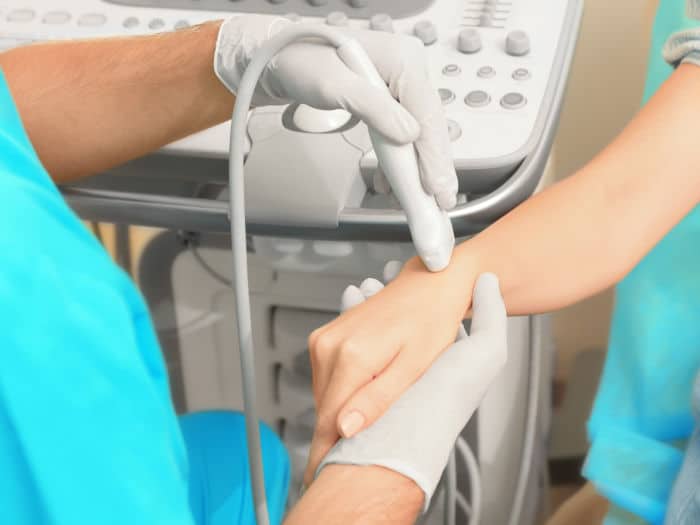
Our Lumps and Bumps Ultrasound Scan
At Universal Ultrasound Diagnostics, we are dedicated to providing exceptional private lumps & bumps ultrasound scans at our lumps & bumps ultrasound clinic in Birmingham, UK. We offer flexible and convenient dates and times that suit your needs and preferences. With our cutting-edge Ultrasound systems and highly skilled, qualified and experienced staff, we deliver precise and reliable imaging for both our private clients and referring practitioners in a comfortable and discreet setting. Our commitment to utilising the latest technology ensures we consistently deliver the highest quality performance you can trust.
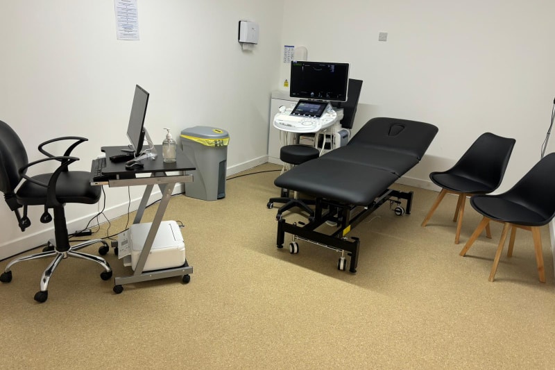
Lumps and Bumps Scan Time & Cost
Typical Cost
You can see our latest rates on our booking page.
Scan Time
Ultrasound scans may take 30 - 50 minutes, depending on the circumstances.
Results
The sonographer will discuss any immediate findings with you, and the report and images will be emailed to you on the same day.
*Polite notice. With Lumps and Bumps, our rate is from these guide prices; if you need more than one area scanned or have several lumps and bumps, we will need to quote you accordingly. Please call us at 0121 392 3112 before booking.
How do we diagnose a lump?
Most of the lumps & bumps that we see in the clinic are benign; however, it is crucial for the sonographer or radiologist to examine them thoroughly. We follow the national recommended guidelines for evaluating lumps and grading them.
Some key steps to getting answers fast are:
- Listening & Examination: It's vitally important for our sonographer, with your permission, to examine and palpate the area or concern/lump after taking your brief medical history (including symptoms, how long the lump has been there, any changes in size, pain, infection or family history of cancer).
- High-resolution ultrasound: Our real-time lumps and bumps scanning in the West Midlands can determine the lump’s precise dimensions, depth, and exact location (such as under the skin, fat, muscle, or gland, or if it is a lymph node). We can determine whether a lump is solid, cystic (filled with fluid), a lipoma (fatty), inflammatory, or post-traumatic via our private ultrasounds. Doppler is used to assess the blood flow and your sonographer or radiologist will analyse the lump to determine if it is benign or malignant.
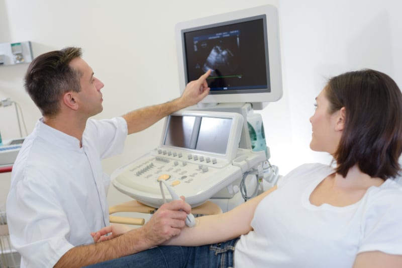
- Quick explanation: Results are reviewed with you right after the scan, and you will receive a report and images via email on the same day. We advise that you share your report with your GP.
- Next steps, if required: If the appearance of the lump is indeterminate or suspicious, we will recommend further testing through your general practitioner to reach a diagnosis, as ultrasound alone cannot diagnose an illness.
Our Mission, Vision And Values
Mission - To be the most efficient and preferred diagnostic imaging centre that provides excellent quality care, that not only meets but goes above and beyond to exceed patients’ expectations.
Vision - Our vision is innovation as healthcare and technology are continuously evolving, which will enhance scanning techniques, image quality, accuracy, and efficiency and provide clearer insights into diseases.
We are proud to be a compassionate, caring, and respectful team. We do this by asking staff to work within our core values:

Compassionate by being inclusive, attentive, caring, respectful and showing kindness to staff, our patients, and their families.

Caring by going beyond compliance, performing above and beyond to improve our services and providing person-centred care by involving patients in their care, offering choices, and promoting dignity and privacy.

Respectful by active listening, seeking to understand others, showing empathy for differences, apologising when things go wrong, being polite and showing gratitude to our staff and patients.

Why Choose Our Private Ultrasound Scan Clinic?
1. Expert Sonographers: Our clinic boasts highly skilled and experienced sonographers specialising in Diagnostics and Pregnancy scans.
2. State-of-the-Art Technology: We use cutting-edge ultrasound systems for precise and reliable imaging.
3. Comfortable and Discreet Setting: Our clinic ensures a comfortable and confidential patient environment.
4. Flexible Appointments: We offer flexible dates and times to suit your convenience.
5. Prompt Results: Immediate findings are discussed during your appointment, and a detailed report and images are emailed to you the same day.
Meet the Team
Lydia, our CQC Registered Manager and Operations Director, leads the team with empathy, faith and the utmost integrity. Her patient-focused approach helps to shape the culture of our service.
Our Consultant Radiologist Dr Moti, is supported by our advanced sonographer Andrew, combine clinical excellence with reassurance, giving our patients peace of mind and genuine warmth. Ali brings clarity to every scan and calm assurance, helping patients feel at ease, supported, and well-informed.
Behind the scenes, Emma, our practice manager, plays a pivotal role, ensuring every detail of the patients’ journey is seamless. She works alongside the Registered Manager to ensure quality assurance is maintained.
Carly, our Medical Administrative Officer, plays a crucial role in patient experience and works alongside the Registered Manager to maintain high-quality standards through promoting continuous improvement across the practice.
Together as a team, we are united by one purpose:
Our Mission - To deliver high-quality care with compassion that not only meets the patients’ expectations but goes over and beyond to exceed expectations.
It's easy to book an appointment.
Universal Ultrasound Diagnostics offers an online booking system for easy appointment scheduling. There are options for walk-ins depending on availability: urgent scans within 5 days and routine scans within 14 days.
Appointment notifications and reminders are sent via email. The system supports self-check-in and online referrals by GPs.
We provide chaperones and interpreting services and allow family presence during pregnancy scans.
Our facility is accessible by public transport and has parking.
We allow family presence for most scans; however, children are not allowed in the room for certain scans. Please check your appointment confirmation email for clarity on our safety policy.
Our FAQ page answers a lot of general questions about your scan.
There is parking around The Green or Pay & Display at the side of The Bulls Head Pub, which charges £1 per hour (cash and card accepted). We are open 8 am to 11 pm Monday to Sunday.
Opening Times
Monday to Sunday 8 am - 11 pm
Location
10c The Green, Kings Norton, Birmingham, B38 8SD

Lumps and Bumps Scan Faqs
ARE LUMPS AND BUMPS NORMAL?
Most lumps are normal. Most people may get a lump or growth on their skin at some point.
A lump and bump can:
• Be hard or soft to touch
• Move around
• Be the size of a pea or a golf ball
• Be a lump under the skin or a growth that hangs off your skin
Lumps and Growths
Lumps can appear anywhere on your body. Most lumps are harmless, but it is important to see a GP if you are worried or the lump is still there after 2 weeks. Alternatively, if you are concerned and do not wish to wait to be seen via the NHS, you can self-refer for an ultrasound scan with us.
Things to be concerned about are: if the lump
- gets bigger
- is painful, red or hot
- is hard and does not move
- lasts more than 2 weeks
- grows back after it has been removed
- is on the groin and does not go down
If you have a lump or bump on or in your breast, testicles or an area of your neck, then you may need a specific soft tissue scan, such as the Breast ultrasound, Scrotal Testicular ultrasound, or Thyroid ultrasound, which we also offer. If you are unsure, please contact your GP or give us a call to discuss your concerns.
A lump and bump can be sebaceous cysts, which are slightly hardened, fluid-filled bumps within the skin, and these are usually considered to be noncancerous. The size of a sebaceous cyst ranges from pea-sized, at about one centimetre across, to around five centimetres across; however, it is advisable to seek medical help.
WHAT ARE THE SYMPTOMS OF COMMON LUMPS AND BUMPS?
Usually, a lump or bump that you find on the body is caused by a small pocket of fluid called a cyst. These are usually not painful nor harmful however. it is best to monitor them as they can grow. Most small cysts will resolve on their own; however, some larger cysts may require draining if they become uncomfortable.
Common areas where cysts can be found on the body include:
• Scalp
• Face
• Neck
• Back
These cysts are usually harmless; however, you should see your GP if you have any of the following symptoms associated with a cyst:
• The diameter is larger than five centimetres
• Immediate recurrence in a very short span of time after removal
• If the cyst is painful, hot to the touch or has a rash or any kind of leakage
In these cases, your GP may advise you to have the lump removed and tested for infection and other causes of illness. Your GP will advise accordingly.
WHAT KIND OF THINGS CAN A LUMPS AND BUMPS ULTRASOUND DETECT?
An ultrasound scan will be able to give a detailed view of the inside of the lump. Looking at the texture will allow the sonographer to see if the lump is a cyst or a tumour; they will also be able to see if it is caused by infection.
Ultrasound cannot diagnose illness alone, and the sonographer may advise you to speak to your GP to be referred for further testing if they are concerned by the images on the scan.
Further testing may involve another type of scan, such as a CT, a punch biopsy to test if the lump is cancerous, or surgery to remove the lump altogether. If you are concerned about any further testing, you should speak to your GP.
Get in touch to book a Lumps and Bumps Scan in the West Midlands
WHAT ARE THE POTENTIAL CAUSES OF A LUMP OR BUMP?
Blocked sebaceous glands, swollen hair follicles, high levels of testosterone, use of androgenic anabolic steroids and trauma are some of the causes of cysts. You may not even have realised that you have caused any trauma to the area, for example, you may develop a lump on your lower back if you wear a rucksack regularly. Not everyone will develop a cyst from such a repeating action. Sometimes cysts can have hereditary causes as well.
What are blocked sebaceous glands?
The sebaceous gland produces the oil called sebum that coats the hair and skin. Cysts can develop if the gland or its duct from which the oil is able to leave becomes damaged or blocked.
What are swollen hair follicles?
A swollen hair follicle or folliculitis is a skin condition in which hair follicles become inflamed. Bacterial or fungal infections are the main culprits of folliculitis. At first, it looks like small red bumps or white-headed pimples around hair follicles, which are the tiny pockets from which hair grows.
What are high levels of Testosterone?
Testosterone is the most important androgen in the human body. Androgens encourage sebum production. Therefore, due to higher androgen levels, seborrheic glands produce more sebum, which is an oily substance leading to coarse pores of the skin. Consequently, acne and acneiform eruptions are more common in seborrheic areas such as the face, upper thorax, back, and shoulders. Due to androgen, there is abnormal thickening of the outer layer of the skin and the sebum is trapped within the follicle and this, in turn, leads to acne lesions and also causes cysts.
What is the use of androgenic anabolic steroids?
High dosages of anabolic-androgenic steroids increase skin surface lipids, and these lipids cover the outer layer of the skin and cause Propionibacteria acne and sebaceous cysts.
What are hereditary causes?
Gardner syndrome is a medical condition where GI polyps, multiple osteomas, and skin and soft tissue tumours are formed. The skin symptoms associated with Gardner syndrome include epidermoid cysts, desmoid tumours, and other benign tumours. Basal cell nevus syndrome is a combination of health defects, which is caused by a rare genetic condition. This condition affects the skin, endocrine system, nervous system, eyes, and bones.
WHAT DO CYSTS USUALLY CONTAIN?
Cysts are generally mobile masses that consist of:
• Fibrous tissues and fluids.
• A fatty (keratinous) substance that resembles cottage cheese and due to this the cyst may be called keratin cyst. This material has a cheesy appearance to it as well as emits a foul odour.
• It may sometimes contain serosanguineous fluid. A serosanguineous fluid generally contains purulent and bloody material.
HOW DO I PREPARE FOR A LUMPS AND BUMPS SCAN?
You will be able to eat and drink as normal before your scan and also take any medications you are on. Please refrain from using any cream or lotion on the area as this may affect the gel needed to aid the scan.
WHAT DO I WEAR?
You will need to loose and comfortable clothing. You will need to expose the area being scanned for the sonographer. If the lump is in an intimate area then the sonographer will do their best to make sure your dignity is maintained throughout the scan.
WHAT DOES THE ULTRASOUND EQUIPMENT LOOK LIKE?
The ultrasound machine consists of a computer console, video monitor and an attached probe called a transducer. The probe uses high-frequency sound waves to build a picture of the inside of the body.
HOW DOES THE PROCEDURE WORK?
The high-frequency sound waves “bounce” back of organs and tissues creating an echo effect that the transducer then translates into the image seen on the screen. The pitch, direction and time it takes the waves to reach the probe all help to determine what the picture should look like.
WHAT ARE THE RED AND BLUE BLOBS ON THE ULTRASOUND?
During the scan, the sonographer may use a colour doppler to check blood flow in your tissue and organs. The colours will appear as blue and red blobs on the image and are a display of real-time sound waves detected in the blood vessels. They are nothing to be concerned about.
HOW IS THE PROCEDURE PERFORMED?
You will be asked to lie on the exam couch and expose the area where the lump or bump is located. A gel will be applied to the skin and the sonographer will move a probe through it and across the area being examined. During the scan, the sonographer may ask you to change position to sit or stand so they can examine the area when you are in different positions. Do not be alarmed if they look at the same area multiple times or go quiet. This is usually nothing to be concerned about as the sonographer needs to concentrate and make sure they have seen and assessed all parts of the area. If there are any concerning findings the sonographer will talk you through what they see and recommend a follow-up.
WHAT WILL MY CHILD EXPERIENCE DURING AND AFTER THE PROCEDURE?
As above your child will be asked to lie on the exam couch and gel applied to their skin. The scan itself should be painless (unless there are any tender areas) and will be fairly quick and easily tolerated. During the procedure, the sonographer may need to check blood flow with the colour Doppler. While this is in use they may hear pulse-like sounds but should not be alarmed by this. The sonographer will warn them if there are going to be any noises. Once the scan is complete the gel will be wiped off your child’s skin and will have no lasting effects. The child will then be able to resume their normal activities. Any findings on the scan will be discussed with you directly.
DO I NEED TO CONSENT TO A LUMPS AND BUMPS SCAN?
All patients must give their consent (permission) before undergoing an examination. If they are unable to give consent themselves then a person with power of attorney will need to give consent on their behalf. Please ensure that the pre-treatment consent form is completed via the embedded link in your confirmation email before you attend the appointment. The scan will not go ahead if this form is not completed.
ARE LUMPS AND BUMPS SCANS PAINFUL?
Ultrasound scans of lumps and bumps are not painful but they may be uncomfortable as the sonographer will have to gently press the ultrasound scan probe to the affected area. If the area is tender this may cause some discomfort.
WHAT ARE THE LIMITATIONS OF ULTRASOUND?
Ultrasound waves are disrupted by air and gas and therefore ultrasound scans are not ideal for imaging air-filled bowel, or organs that may be obstructed by the bowel. Ultrasound cannot penetrate areas hidden by bone such as head or lungs and therefore cannot be used to diagnose fractures or breaks.
Larger patients may also be more difficult to scan using ultrasound because of the greater amounts of tissue. This can weaken the sound waves and thus affect the images.