


Venous insufficiency
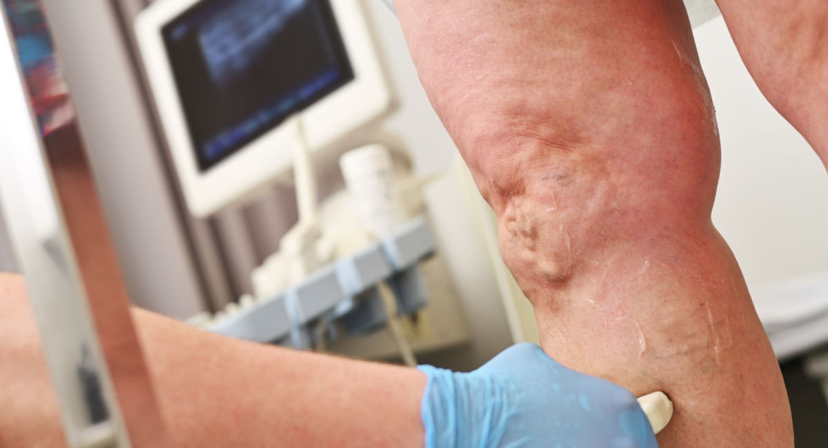
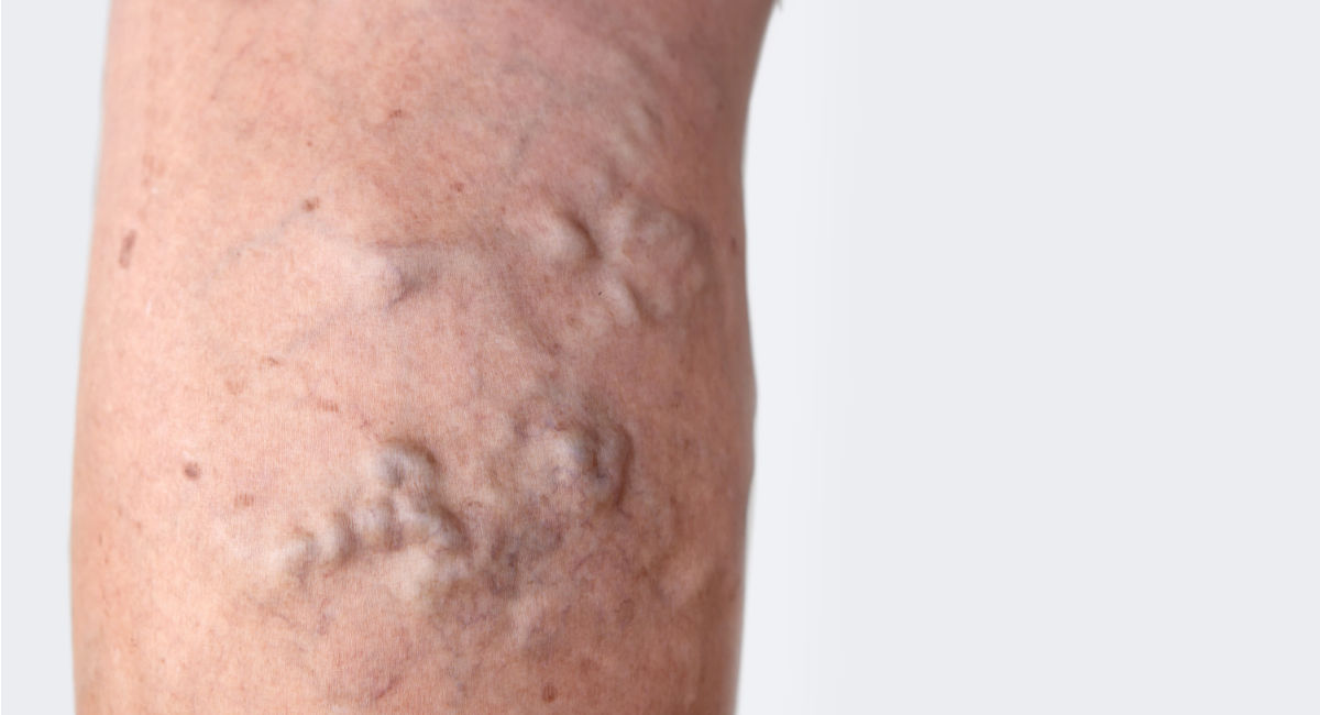
If you have aching legs, ankle swelling that builds through the day, visible veins, or skin changes around your lower legs, you may have heard the term venous insufficiency (also called chronic venous insufficiency or chronic venous disease).
It is common, it can be uncomfortable, and when it is ignored for years, it can lead to skin damage and, in more advanced cases, venous leg ulcers. The good news is that there are practical steps you can take to reduce risk and slow progression, and there is a clear, evidence-based pathway for diagnosis and management.
This guide explains the science in plain English, including how Venous Insufficiency ultrasound (duplex scanning) is used to confirm the diagnosis, map the problem veins, and guide further clinical correlation and treatment decisions.
What is Venous Insufficiency?
Your leg veins carry blood back to the heart against gravity. To do that efficiently, veins rely on one-way valves inside the veins that stop blood from falling backwards, the calf muscle pump (walking and ankle movement squeeze the deep veins and push blood upward), and healthy vein walls that help prevent pooling.
Venous insufficiency happens when this system becomes less effective, most often because the valves do not close properly (valve incompetence). When valves fail, blood can leak backwards (reflux) and pool in the lower legs. Over time, this increases pressure in the veins, which can lead to symptoms like heaviness and swelling, and later skin changes and ulcers.
Venous insufficiency can involve superficial veins (often linked with varicose veins), deep veins, perforator veins (which connect superficial and deep systems), and sometimes central veins in the pelvis or abdomen if there is obstruction upstream.
Common causes and risk factors
In our clinic, patients usually ask what is causing their leg symptoms and if they have venous insufficiency or something else.
Venous insufficiency is usually multifactorial, meaning several factors combine.
Common contributors include:
- Prolonged standing or sitting (reduced calf pump activity)
- Age and reduced mobility
- Pregnancy and hormonal factors
- Excess weight
- Family history
- Previous deep vein thrombosis (DVT), which can damage valves or disrupt flow
Prevention and self-care
While there is no single prevention step, the evidence supports reducing venous pressure and improving circulation.
Keep the calf pump working:
- Walk regularly, even short, frequent walks
- Add ankle movements if you are desk-based
- Break up long sitting or standing periods
Support a healthy weight:
Weight management reduces strain on leg veins.
Elevate your legs when you can:
Leg elevation can reduce swelling and discomfort, particularly later in the day.
Look after your skin:
If you have dry, itchy or scaly skin on the lower legs, moisturise regularly. Skin breakdown increases the risk of complications.
Compression, when appropriate:
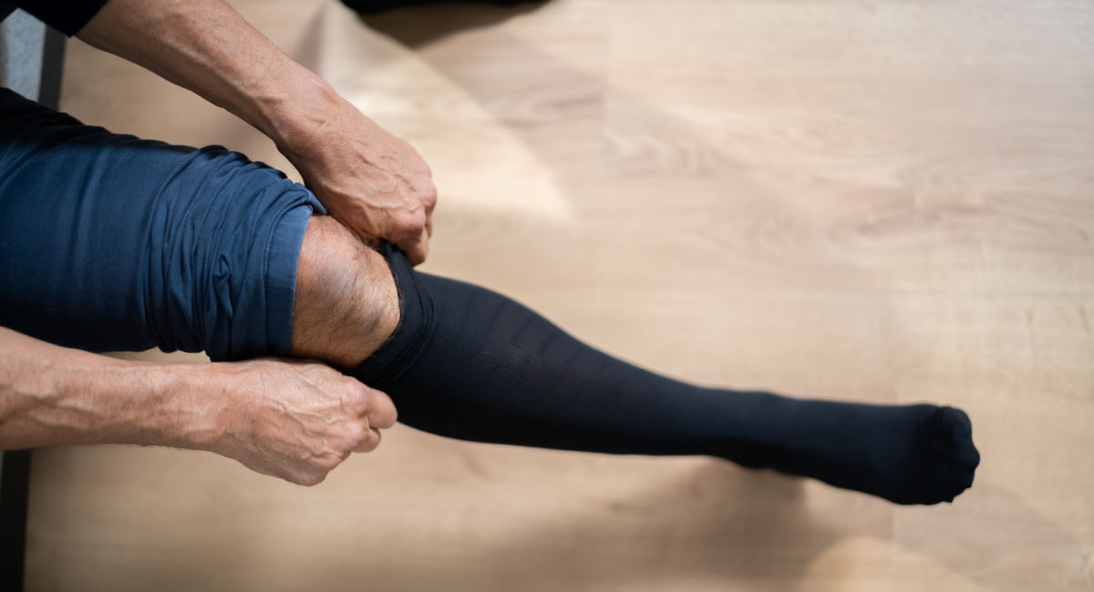
Compression can reduce swelling and support venous return. It is a mainstay of treatment for venous ulcer care and is often used for symptom control. Compression should be selected carefully, particularly if there is any concern about arterial circulation.
How venous insufficiency is diagnosed
When patients call us to find out if their skin changes or symptoms can be explained by a venous insufficiency scan, we take their history to help them book for the right scan.
Step 1: History
At our clinic, our advanced sonographer will ask about symptom pattern, triggers, previous clots, surgery, pregnancy history, and any skin changes or wounds. The sonographer will examine the legs for varicose veins, swelling, skin changes and areas of tenderness.
Step 2: Duplex ultrasound (the key test)
Our sonographer will explain the examination to the patient, informing them that an ultrasound scan will help to understand which veins are affected (superficial, deep, perforators), whether there is reflux, obstruction, or both, and how severe it is. After the sonographer will explain findings and advise the patient to share their report with their doctor (GP), who can discuss with them an appropriate management or refer them to a vascular specialist.
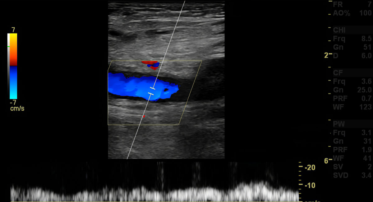
Duplex ultrasound combines B-mode imaging (a picture of the vein) and Doppler flow assessment (how blood is moving, direction and speed). NICE states that people referred to a vascular service for varicose veins should have a duplex ultrasound scan because it identifies blood flow patterns and helps locate damaged valves. International guidance also supports duplex ultrasound as the diagnostic test of choice in chronic venous disease.

The role of ultrasound in diagnosis and management
Ultrasound is not just a yes-or-no test. It provides a detailed map that can help to guide change management.
1) Confirming reflux (valve failure)
A reflux study looks for backward flow in key veins, often while standing and using specific manoeuvres. It helps identify which superficial veins are incompetent (for example, the great saphenous vein), whether perforator veins are contributing, and whether deep venous reflux is present.
2) Checking for obstruction, including deeper or central issues
Some people have venous outflow obstruction, including iliac vein compression or post-thrombotic narrowing. Guidance notes that in suspected more proximal obstruction, assessment may include abdominal or pelvic venous ultrasound in addition to full leg duplex assessment.
3) Ruling out DVT when symptoms suggest a clot
Venous insufficiency and DVT can overlap in symptoms like swelling and pain. If a DVT is clinically suspected, rapid ultrasound is part of the recommended pathways.
If you ever have leg swelling or pain plus breathlessness or chest pain, treat that as an emergency and seek urgent care.
4) Planning treatment (mapping)
If a patient is suitable for procedures such as endovenous ablation or sclerotherapy, ultrasound mapping helps identify the faulty segment accurately and therefore supports safer, more targeted treatment decisions.
5) Monitoring outcomes
An ultrasound can also be used to check for treatment success, complications, and track progression in complex cases.

What happens during a venous duplex ultrasound?
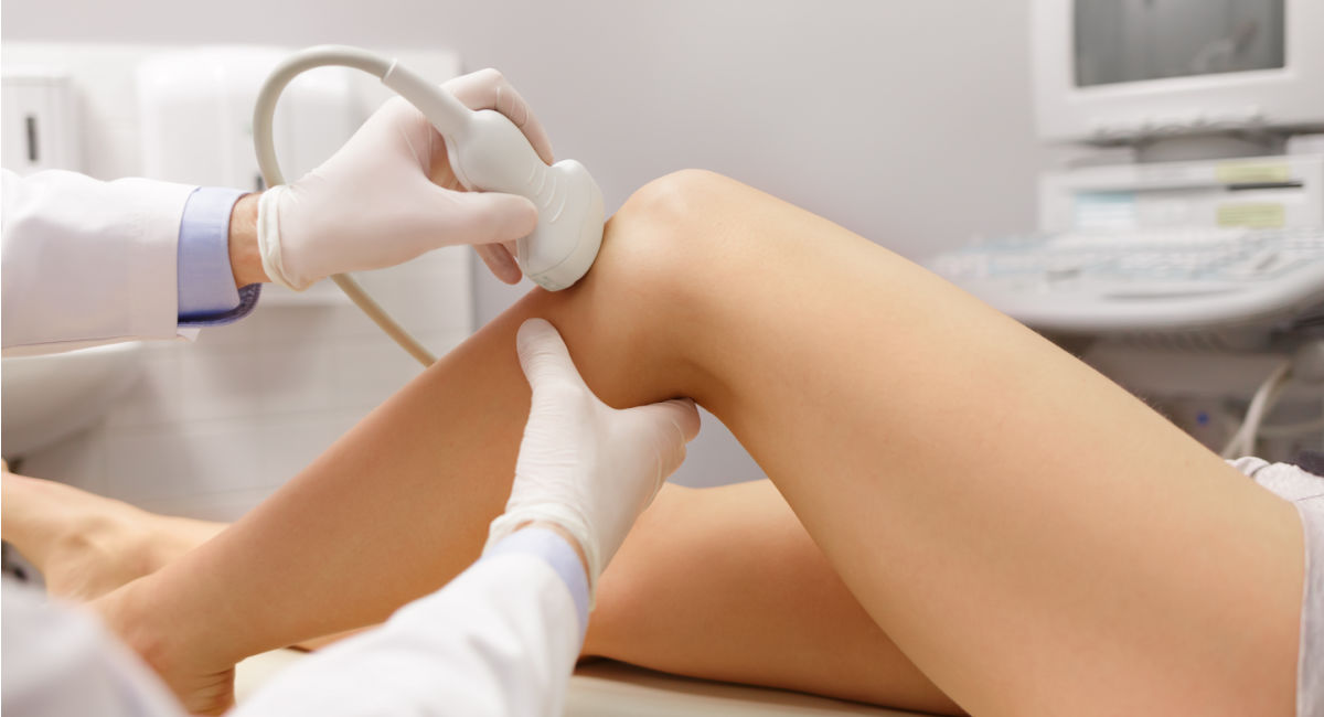
Most venous duplex scans are straightforward. The sonographer uses gel and a probe to visualise veins along the leg and Doppler to assess flow direction and valve function. Depending on the purpose, you may be scanned lying down and standing. The scan is non-invasive and uses sound waves, not radiation.
Severity classification (CEAP)
Specialists often describe chronic venous disease using the CEAP classification, which ranges from no visible disease (C0) to active ulcers (C6).
Treatment and management: what typically helps
Management depends on severity, symptoms and ultrasound findings. Common recommendations include lifestyle measures (walking, calf strengthening, weight management, leg elevation, skin care) and compression where appropriate. For venous ulcer care, compression is central to improving venous return and reducing swelling.
If symptoms affect quality of life, or there are skin changes or ulceration, referral into vascular pathways is often appropriate.
When to seek medical advice urgently
Seek urgent assessment for sudden one-sided leg swelling and pain, redness and warmth in the calf, or any DVT-like symptoms plus breathlessness or chest pain. Also seek help for bleeding from a varicose vein, rapidly worsening skin changes, or an open wound that is not healing.
Where a private ultrasound scan fits in
A private venous insufficiency duplex scan can be helpful when symptoms are persistent, and you want clarity on what is happening in the veins, or when you want a detailed reflux assessment to support next steps. If you have symptoms that suggest DVT or pulmonary embolism, this needs urgent NHS assessment rather than waiting for a routine appointment.
Key Takeaway
Venous insufficiency is a circulation problem that can progress if underlying reflux or obstruction is not identified and managed. Duplex ultrasound is central because it shows where blood is flowing the wrong way, which valves are not working properly, whether there are signs of obstruction or previous clot-related changes, and the pattern that guides safe treatment decisions.
Why choose a private ultrasound scan
Choosing the right private healthcare clinic is all about access, reassurance, and discretion. Our Birmingham private ultrasound clinic is easy to reach, with quick, flexible appointments and a welcoming team dedicated to your comfort. We are a highly rated and trusted Private Ultrasound Clinic. Read our client reviews.
Take the next step today:
- Book your scan online
- Request a call back to discuss the best scan for you
- Message us with any concerns; no question is too small
Contact Ultrasound Diagnostic, Birmingham’s accessible, confidential choice for Venous Insufficiency ultrasound and DVT ultrasound scans. Use our Online Booking System to schedule an ultrasound scan appointment and feel confident about your health.